UNIVERSIDADE FEDERAL DE CIÊNCIAS DA SAÚDE DE PORTO ALEGRE UFCSPA CURSO DE PÓS-GRADUAÇÃO EM CIÊNCIAS DA SAÚDE. José Antonio Tesser Poloni
|
|
|
- João Gabriel Peralta Figueiredo
- 6 Há anos
- Visualizações:
Transcrição
1 UNIVERSIDADE FEDERAL DE CIÊNCIAS DA SAÚDE DE PORTO ALEGRE UFCSPA CURSO DE PÓS-GRADUAÇÃO EM CIÊNCIAS DA SAÚDE José Antonio Tesser Poloni Validação da Técnica de Detecção de Células Decoy por Microscopia Óptica Convencional Porto Alegre
2 José Antonio Tesser Poloni Validação da Técnica de Detecção de Células Decoy por Microscopia Óptica Convencional Dissertação submetida ao Programa de Pós-Graduação em Ciências da Saúde da Fundação Universidade Federal de Ciências da Saúde de Porto Alegre como requisito para a obtenção do grau de Mestre Orientadora: Dra. Liane Nanci Rotta Co-orientadora: Dra. Elizete Keitel Porto Alegre
3 3
4 Ao meu filho Pedro, que está se preparando para o momento mais importante e mais aguardado de nossas vidas! 4
5 AGRADECIMENTOS Agradeço aos meus pais e a minha esposa por todo o amor e apoio que sempre dedicaram, não medindo esforços para dar as melhores condições possíveis para que eu pudesse me dedicar ao meu trabalho e estudos. A minha orientadora e a minha co-orientadora pelo suporte e apoio recebidos durante todo o período do mestrado. As equipes do Laboratório Central de Análises Clínicas ISCMPA, do Laboratório de Biologia Molecular ISCMPA, do Laboratório de Micologia e Patologia ISCMPA e do Ambulatório de Nefrologia ISCMPA pela ajuda para que o trabalho fosse possível. Sinceramente, faltam palavras e espaço suficiente para citar nominalmente cada uma das pessoas que me ajudou na condução do projeto. Recebi ajuda de muitos amigos, cada um com sua parcela fundamental de participação. Ninguém alcança objetivo algum sozinho, sem a ajuda de um grande número de pessoas que dão suporte e ajudam das mais diversas formas. Este trabalho só foi possível devido ao esforço de muitas pessoas que abriram mão de momentos de suas vidas para auxiliar um amigo. Jamais conseguirei agradecer o suficiente a todos que colaboraram comigo para que eu pudesse concluir este trabalho. 5
6 RESUMO O poliomavírus BK (BKV) é um agente patogênico que está fortemente associado à perda do enxerto em pacientes transplantados renais e é de grande prevalência na população. Os métodos diagnósticos utilizados para detecção do BKV ou mais especificamente das células decoy (células epiteliais tubulares renais modificadas pelo BKV) são trabalhosos, onerosos e muitas vezes requerem procedimentos invasivos, como por exemplo, a biópsia renal. A análise do sedimento urinário por contraste de fase já se mostrou eficiente para detecção destas células, porém, este recurso é muito pouco utilizado em laboratórios de análises clínicas. A análise do sedimento urinário por microscopia óptica convencional (campo claro) também pode ser utilizada para identificação destas células. O objetivo deste trabalho foi padronizar a técnica de detecção das células decoy utilizando microscopia de campo claro, a fim de introduzir este elemento na gama de achados reportados no exame comum de urina. Os resultados obtidos com este trabalho evidenciaram concordância entre os achados de células decoy por microscopia de campo claro e por coloração de Papanicolaou (P<0,001). Também houve concordância do achado de células decoy com a virúria (P<0,001) e a viremia por BKV (P<0,001). Sensibilidade e especificidade da microscopia de campo claro, quando comparado ao padrão-ouro (imunoistoquímica da biópsia renal) foi 80% e 52%, respectivamente. Conclui-se que é possível realizar a identificação segura e confiável das células decoy no sedimento urinário sem utilizar qualquer tipo de coloração e com recurso de microscopia de campo claro, que está amplamente disponível em todos os laboratórios de análises clínicas. Este achado permite que se forneça de maneira rápida, indolor e com baixo custo a informação de uma possível reativação do BKV. Palavras-chave: Células decoy, Poliomavírus BK, Sedimento urinário, Microscopia de campo claro 6
7 ABSTRACT The polyomavirus BK (BKV) is a pathogenic agent strongly associated with graft loss in renal transplant recipients and have high prevalence in the population. The diagnostic methods used for detection of BKV or more specifically the decoy cells (renal tubular epithelial cells modified by the BKV) are laborious, expensive and often require invasive procedures, such as kidney biopsy. The analysis of urine sediment by phase contrast has proved efficient for the detection of these cells, however, this feature is rarely used in clinical laboratories. The analysis of fresh and unstained urinary sediment by bright field microscopy can be used to identify these cells. The objective of this study was to standardize the detection of decoy cells using bright field microscopy in order to introduce this find on the elements that can be reported during routine urine sediment analysis. The results obtained in this study revealed agreement between the finding of decoy cells by bright field microscopy in the fresh urine sediment and by Papanicolaou staining (P<0.001). There was also agreement between the finding of decoy cells with BKV viruria (P<0.001) and BKV viremia (P< 0.001). Bright field microscopy sensitivity and specificity, compared to gold standard (immunohistochemistry of the kidney biopsy) were 80% and 52%, respectively. Thus, we can conclude that it is possible to perform safe and reliable identification of decoy cells in urine sediment without using any kind of stain and using bright field microscopy, which is widely available in all clinical laboratories. This finding allows to provide quick, painless and inexpensive information of a possible reactivation of BKV. Keywords: Decoy cells, Polyomavirus BK, Urine sediment, Bright field microscopy 7
8 LISTA DE ABREVIATURAS BKV: BK vírus DC: Decoy cells (células decoy) DC BFM: Decoy Cell under Bright Field Microscopy EQU: Exame Qualitativo de Urina HIV: Human immunodeficiency virus JCV: JC vírus MMF: Micofenolato mofetil PCR: Polimerase chain reaction PVN: Polyomavirus nephropathy SV40: Simian virus 40 8
9 SUMÁRIO CAPÍTULO I REVISÃO DA LITERATURA 10 1 Introdução Poliomavírus Nefropatia por Poliomavírus (PVN) Fatores de risco Diagnóstico da PVN Pesquisa de células decoy Tratamento Prognóstico Justificativa Objetivos Objetivo geral Objetivos específicos Referências bibliográficas...23 CAPÍTULO II - Artigo científico...27 Conclusão...44 Anexos
10 Capítulo I REVISÃO DA LITERATURA 10
11 1 INTRODUÇÃO 1.1 Poliomavírus A família Polyomaviridae inclui três membros: o JC vírus (JCV), o simian vírus 40 (SV40) e o BK vírus (BKV) [1,2]. Tratam-se de vírus com estrutura molecular muito semelhante, medindo 40 nm de diâmetro e que possuem genoma circular de 5,1-kb, composto de DNA dupla fita. Enquanto o SV40 é comumente encontrado em macacos, sendo de pouco significado em humanos, o JCV é um importante causador de doença neurológica em pacientes infectados pelo vírus da imunodeficiência humana (HIV), agente de leucoencefalopatia progressiva multifocal [3]. Diferentemente do BKV, nenhum dos demais poliomavírus têm sido associados de modo consistente com nefropatia pós-transplante renal. As primeiras poliomaviroses, causadas por BKV e por JCV, foram coincidentemente isoladas em 1971 por dois grupos independentes: o BKV em urina de um paciente transplantado renal e o JCV do tecido cerebral de um paciente com linfoma de Hodking [4]. Classicamente, a infecção primária por BKV acontece na infância, sendo muito comum, variando desde casos assintomáticos até sintomas sugestivos de um simples resfriado [5,6]. A transmissão ocorre primariamente por via oral ou respiratória, sendo também possível adquirir o vírus através de transfusões sanguíneas, transplantes ou mesmo por via transplacentária [7]. Após a infecção primária, em indivíduos imunocompetentes, o vírus entra em um período de latência com tropismo pelo trato urinário (células tubulares renais, cápsula de Bowman e células uroteliais) [5,6,8]. Recentemente foi evidenciada uma associação entre a presença de hematúria assintomática e infecção urinária por poliomavírus em pacientes imunocompetentes [9]. A soroprevalência para o BKV e para o JCV nos adultos é muito alta: mais de 90% da população adulta é soropositiva para o BKV, enquanto que 50 a 80% dos adultos tem anticorpos para o JCV. Em indivíduos imunocompetentes não causam patologia, porém, em indivíduos imunocomprometidos as poliomaviroses humanas causam doença primária significante, permitindo a reativação de um estado subclínico persistente para 11
12 uma infecção lítica, resultando em virúria e viremia, potencialmente podendo conduzir a doença severa ou fatal [4]. Para o BKV, a reativação é mais comum em pacientes que receberam transplante de medula e em pacientes que receberam transplante renal, onde a infecção lítica do BKV resulta em cistite hemorrágica e em nefropatia por poliomavírus, respectivamente. Ano passado, pela primeira vez foi reportado que a replicação do BKV nos pacientes receptores de transplante renal tem origem no doador. A urina de 249 pares doador/receptor foi investigada para a presença de DNA de BKV por qpcr antes do transplante e consecutivamente depois do transplante [10]. A reativação tem sido observada em indivíduos com condições imunes alteradas, incluindo pacientes com HIV [4], transplantados de órgãos sólidos [11] e doenças autoimunes como lúpus eritematoso sistêmico. Esta última condição foi referenciada pelo nosso grupo, sendo demonstrada pela detecção das células decoy, pela primeira vez na literatura médica, e comprovada através da reação em cadeia da polimerase (PCR) [12]. 1.2 Nefropatia por poliomavírus (PVN) A PVN tem se tornado um problema emergente na população de receptores de transplante renal [13,14]. Com prevalência elevada e sem tratamento específico, pode levar à perda precoce do enxerto com grande morbidade, fazendo com que o paciente retorne a programas de terapia de substituição renal, o que eleva os custos do tratamento. Isto tem sido motivo de estudo em centros transplantadores de todo o mundo. A virúria e a viremia causadas por BKV, decorrentes da reativação, são encontradas em 80% dos pacientes de transplante renal e 10% destes pacientes progridem para PVN, resultando em perda do enxerto em 90% das vezes [4]. A PVN é caracterizada por necrose dos túbulos proximais e desnudação da membrana basal como resultado da ação lítica da infecção pelo BKV nas células epiteliais renais [4]. Com a introdução de novos e mais potentes regimes de imunossupressão, a incidência de PVN tem aumentado [4], indicando uma relação entre a reativação do BKV e o rompimento do sistema imune. A prevalência de nefropatia associada ao BKV está em torno de 2-8% [15-19]. Após o diagnóstico da PVN, cerca de 50% dos pacientes evoluem para 12
13 perda progressiva de função do enxerto renal em 2 anos [16-20], com necessidade de retorno à terapia renal substitutiva. Um estudo retrospectivo e comparativo demonstrou sobrevida média do enxerto de 47,8 meses e 88,7 meses, respectivamente, para pacientes com e sem presença de PVN [16]. Na ausência de intervenções, a perda do enxerto é evidenciada em 45% dos casos após 6 meses do diagnóstico [21]. O diagnóstico precoce da replicação viral permite intervenção terapêutica com redução do risco de desenvolvimento da doença e, por conseguinte, da perda de enxerto [18,20,22]. Desde 1995 que a PVN vem sendo reconhecida como uma causa significativa de disfunção dos rins transplantados com consequente perda do enxerto. Ela em geral ocorre de 2 a 60 meses depois do transplante, mas é mais frequentemente identificada dentro do primeiro ano pós-transplante. 1.3 Fatores de risco Os fatores de risco para desenvolvimento de infecção por BKV não são totalmente compreendidos. Enquanto estudo envolvendo crianças demonstrou maior risco de nefropatia por BKV em receptores soronegativos, que receberam rins de doadores soropositivos [23], Hirsch e colaboradores demonstraram que 85% dos pacientes com nefropatia por BKV apresentavam sorologia positiva pré-transplante [15]. Pacientes do sexo masculino e com idade avançada apresentam maior incidência de nefropatia por BKV [16]. O uso de stents ureterais tem sido associado com a ocorrência de virúria persistente por BKV [24] Outros fatores relacionados ao receptor, tais como variáveis demográficas e imunológicas, bem como tempo de isquemia e a função inicial do enxerto, não parecem se associar com risco aumentado para desenvolvimento da nefropatia por BKV [2,25]. A introdução de novos agentes imunossupressores, tais como micofenolato mofetil (MMF) e tacrolimus, tem demonstrado relação temporal com a ocorrência de nefropatia por BKV. Uma incidência aumentada da doença tem sido descrita em pacientes recebendo esquema imunossupressor tríplice com MMF, tacrolimus e prednisona [26], o que não tem sido observado em todos os estudos [25]. Já o uso de anticorpos policlonais para terapia de indução não teve associação com presença de células decoy ou nefropatia por 13
14 BKV [15]. Embora o uso de novas medicações imunossupressoras tenha sido implicado como fator associado ao aumento na incidência de nefropatia por BKV, o principal fator determinante ainda é desconhecido, uma vez que a infecção é diagnosticada em receptores que nunca usaram esquemas imunossupressores potentes, assim como em pacientes em uso de sirolimo e em esquemas livres de corticosteróides [2]. Marcadores não invasivos como as células decoy, virúria e viremia e anticorpos séricos contra o BKV podem ser úteis como marcadores adjuvantes para o diagnóstico da PVN e marcadores para o monitoramento da replicação do BKV e a resposta da PVN a intervenções terapêuticas. Entretanto a redução da imunossupressão deve ser balanceada contra riscos inerentes de rejeição. Em casos onde a rejeição se apresenta simultaneamente com PVN ou ocorre depois do diagnóstico da PVN, um procedimento em duas etapas é recomendado consistindo de um aumento inicial da imunossupressão para tratar o episódio de rejeição, seguido de uma diminuição da imunossupressão. Atualmente, não existem drogas efetivas não nefrotóxicas para o tratamento antiviral para a PVN [27]. 1.4 Diagnóstico da PVN O padrão atual de diagnóstico da PVN inclui a análise de biópsias renais usando histopatologia ou imunohistoquímica, tanto para detectar mudanças citopáticas como resultado de injúria ou de lise das células epiteliais tubulares renais, ou para detectar a expressão genética do vírus. Também é utilizada a avaliação da citologia urinária para detectar a presença de células decoy, que são células epiteliais com corpos de inclusão viral intranuclear, e PCR para detectar os genomas virais e quantificar a liberação viral nas amostras de sangue ou de urina [4]. O PCR e a citologia urinária são especialmente úteis para detectar os estágios iniciais de reativação e PVN, pois permitem diagnósticos e intervenções rápidas, com um consequente aumento da sobrevida do transplantado [4]. A técnica do PCR em tempo real permite diagnosticar e quantificar genomas de BKV em células humanas de sangue periférico de maneira rápida e acurada, com alta sensibilidade e especificidade [6], de forma não invasiva, 14
15 sendo um novo instrumento no diagnóstico de PVN. Ainda hoje, tal metodologia possui custo elevado para sua inserção em massa como rotina diagnóstica na suspeita de infecção por BKV. Embora os testes moleculares sejam hoje mais amplamente disponíveis, é necessário que se determinem os valores de ponto de corte utilizados para predizer o risco de desenvolvimento de nefropatia associada ao vírus, indicando o melhor momento para o início do tratamento da infecção antes que se instale dano tecidual renal irreversível. Em um estudo piloto, foi demonstrado que virúria e viremia por BKV foram documentados em 62% e 42% dos pacientes transplantados renais evidenciando disfunção do enxerto renal [28]. Neste estudo, que empregou técnica de PCR semi-nested, a presença (qualitativa) de virúria intensa (+++) foi associada com maior probabilidade de viremia por BKV. Em trabalho realizado por nosso grupo testando dois protocolos diferentes de extração de DNA viral, um analisando a amostra de urina total e outro analisando o sedimento urinário observamos que existiu uma diferença estatisticamente significativa na quantidade de DNA viral detectada no sedimento urinário, comparado com a urina total, o que demostra uma maior capacidade de detecção do patógeno quando se utiliza este procedimento pré-analítico [29]. Uma vez que testes quantitativos de PCR em tempo real se encontram agora disponíveis, é preciso definir com maior exatidão o ponto de corte de virúria e de viremia que possam predizer a presença de nefropatia por BKV. A detecção de virúria através do método qualitativo de PCR é observada em 30-40% dos receptores de transplante renal [15], sendo um exame com excelente valor preditivo negativo, mas com valor preditivo positivo baixo (40%); é considerado também, um bom teste de rastreamento [2]. A detecção da presença de BKV no plasma, utilizando técnica de PCR é útil ao diagnóstico, precedendo a ocorrência de PVN [2,7]. A análise por imunohistoquímica pode ser realizada em secções de tecido obtidos de biópsia, fixados em formalina e conservados em parafina usando anticorpos comercialmente disponíveis para detecção do antígeno SV40 que é comum para o JCV, BKV e SV40 [30]. A PVN desenvolve-se, em geral, no final do primeiro ano do transplante, com média entre meses [15,16,25]. A presença do vírus na urina, demonstrada por citologia urinária (observação de células decoy) e do DNA viral, precede em aproximadamente 4 semanas o diagnóstico da viremia e 15
16 em 12 semanas a confirmação histológica [15]. Portanto, métodos diagnósticos que permitam a identificação precoce de pacientes em risco de desenvolver doença por BKV devem ser utilizados, devido à possibilidade da administração de intervenções terapêuticas precoces que visem à manutenção da função do enxerto. 1.5 Pesquisa de células decoy A presença de células decoy pode ser um achado de relevância diagnóstica para indicar a reativação do poliomavírus. Usualmente as mesmas são identificadas por coloração de Papanicolaou em urina fixada, tanto em esfregaços de amostra total quanto em amostras citocentrifugadas [19,31]. Em estudo recente foi evidenciado que a pesquisa de células decoy através da citologia urinária demonstrou ser um método não invasivo de grande utilidade para o diagnóstico de nefropatia pelo poliomavírus BK, chegando a encontrar especificidade de 99,7% [32]. Os resultados obtidos em um trabalho publicado por pesquisadores da UFCSPA, conduzido na Irmandade da Santa Casa de Misericórdia de Porto Alegre, indicaram 84,6% de sensibilidade para as células decoy quando comparadas com os resultados das biópsias renais [33]. Pela coloração de Papanicolaou (Figura 1), muitas células decoy mostram um núcleo muito aumentado, que é ocupado por uma inclusão basofílica rodeada pela cromatina, que confere uma aparência de campo vítreo ou gelatinosa. Algumas vezes a inclusão nuclear tem um aspecto vesicular, ou pode ser rodeada por um halo, e a cromatina estar aderida a ele [19,31]. Quando as células decoy derivam do uroepitélio, um núcleo muito aumentado e alterado, assim como a forma irregular do corpo da célula podem mimetizar as mudanças observadas em células neoplásicas [19,31]. Na análise do sedimento urinário por microscopia de contraste de fase (Figura 2) [31], as células decoy mostram as mesmas anormalidades descritas pelos espécimes corados pelo método de Papanicolaou, isto é, aumento do núcleo com uma aparência de campo vítreo ou vesicular, cromatina alterada, nucléolos aumentados, a presença de um halo, e, as vezes, também vacúolos citoplasmáticos. Estas características fazem as células decoy diferentes das 16


.")

17 células epiteliais tubulares e transicionais, encontradas em todas as outras condições. Figura 1 Células decoy coradas por Papanicolaou (400x). Figura 2 Célula decoy vista em microscopia de contraste de fase (400x). Poucos laboratórios de análises clínicas possuem microscópios equipados com filtros para contraste de fase, recurso considerado dispendioso 17
18 na maioria das vezes, devido a pouca valorização que é dada para o Exame Qualitativo de Urina (E.Q.U.) em muitos centros. Pela primeira vez no Brasil, conseguimos comprovar a possibilidade de identificação das células decoy no sedimento urinário de amostras de pacientes que realizam exames no setor de Uroanálise do Laboratório Central de Análises Clínicas da Irmandade da Santa Casa de Misericóridia de Porto Alegre (ISCMPA) e, de maneira inédita na literatura, o fizemos utilizando microscopia óptica de campo claro sem utilizar qualquer coloração [34]. Uma situação de importância para a literatura médica no contexto da nefrologia e mais especificamente da uroanálise, é que antes de 2009 não se encontravam livros com imagens de células decoy em sedimento urinário não corado, fato que começou a mudar neste ano com a publicação do livro do Dr. Giovanni B. Fogazzi The urinary sediment an integrated view [35] onde são apresentadas imagens de células decoy em sedimento urinário vistas por microscopia de contraste de fase. Em 2012, Dr. Fogazzi publicou o livro The urinary sediment by sedimax [36] apresentando imagens das células decoy obtidas através da utilização deste equipamento que realiza análise automatizada do sedimento urinário por obtenção de imagens e de um software de reconhecimento dos elementos. Ano passado Dr. Mark A. Perazella em conjunto com Dr. Robert F. Reilly Jr. publicaram o livro Nephrology in 30 days [37] onde entre outras imagens consta uma de uma célula decoy vista em sedimento urinário não corado através de microscopia de campo claro. Esta imagem que consta neste livro foi obtida no Laboratório Central de Análises Clínicas da Irmandade da Santa Casa de Misericórdia de Porto Alegre. As amostras de urina dos pacientes transplantados renais são um desafio a parte para os microscopistas que atuam em laboratórios de análises clínicas que atendem este perfil de pacientes. São necessárias inúmeras horas de treinamento para que um profissional esteja apto a conduzir uma análise segura que possibilitará, por exemplo, alertar ao médico assistente para uma possível reativação do BKV. A facilidade e simplicidade do exame comum de urina não podem jamais levar o profissional ao relaxamento dos padrões de qualidade necessários para a obtenção de resultados fidedignos. O maior prejuízo que um profissional de análises clínicas que realiza análise de sedimento urinário pode causar é deixar de fornecer uma informação 18
19 clinicamente útil ou classificar determinado elemento erroneamente. O foco principal deste trabalho é apresentar esta célula para estes profissionais que se deparam diariamente com este elemento (Ex.: no Laboratório Central de Análises Clínicas Irmandade da Santa Casa de Misericórdia de Porto Alegre, de 1 a 5 pacientes dos aproximadamente 70 receptores de transplante renal que comparecem aos postos de colheita do laboratório apresentam células decoy nos sedimentos urinários) e precisam ter o conhecimento adequado para fornecer uma informação correta. Desta forma, este tipo de célula é um achado de rotina para os profissionais do Laboratório Central justificando a condução deste trabalho para padronização do procedimento de pesquisa das células decoy. 1.6 Tratamento O manejo da infecção por BKV tem como objetivo reduzir a replicação viral, evitar episódios de rejeição aguda associada à infecção e preservar a função renal. A reversão da imunossupressão é a estratégia utilizada com maior freqüência neste contexto, permitindo ao sistema imune hospedeiro combater a infecção viral [24,18]; tais medidas podem, no entanto, implicar em aumento do risco de rejeição aguda [16]. Sabe-se que a redução da imunossupressão estabiliza a função renal de transplantados com nefropatia por BKV, reforçando-se a importância de se estabelecer um diagnóstico precoce para esta condição [25,38]. O tratamento farmacológico da nefropatia por BKV envolve o uso de drogas que reduzam a carga viral do BKV. O tratamento com leflunomida, associado à redução da imunossupressão, demonstrou importante redução na viremia e virúria [39]. Cidofovir tem sido utilizado no tratamento da infecção; no entanto, este agente é nefrotóxico e excretado pelo rim, necessitando-se maiores cuidados relacionados a hidratação do paciente [40]. Imunoglobulina humana e ciprofloxacino são descritos como opções terapêuticas [41,42]. Estudos prospectivos e randomizados são ainda necessários para que se defina a eficácia das terapias atualmente empregadas para nefropatia por BKV. A prevenção da nefropatia por BKV através do monitoramento da carga viral por PCR e da pesquisa de células decoy em sedimento urinário pode ser uma importante abordagem, permitindo o diagnóstico precoce de pacientes em risco 19
20 para desenvolver a doença e oportunizando intervenção terapêutica antes da lesão renal ser estabelecida, preservando a função do enxerto. Estudos de Brennan e Hussain, com seus respectivos colaboradores, demonstraram que a redução precoce da imunossupressão em pacientes com viremia por BKV foi eficiente e segura em prevenir o aparecimento de nefropatia por este poliomavírus [24,43]. Em crianças, há evidências de que a avaliação seriada de viremia e virúria, com redução da imunossupressão em pacientes virêmicos, foi eficaz em reduzir o risco de desenvolvimento de nefropatia por BKV, sem aumento do risco de rejeição aguda [23]. 1.7 Prognóstico Após o diagnóstico da nefropatia por BKV, em torno de 50% dos pacientes evoluem para perda progressiva de função do enxerto renal, ao longo de dois anos de seguimento [16-20] com necessidade de retorno a terapia renal substitutiva. Um estudo retrospectivo demonstrou sobrevida média do enxerto de 48 meses e 89 meses, respectivamente, para pacientes com e sem presença de nefropatia por BKV [16]. Na ausência de qualquer intervenção, a perda do enxerto é evidenciada em 45% dos casos após seis meses do diagnóstico [21]. A determinação precoce da replicação viral permite intervenção terapêutica, com redução do risco de desenvolvimento da doença e, consequentemente, da perda de enxerto [18,20,23,38]. A possibilidade de fornecer a informação da presença do BKV através da observação das células decoy em um exame simples, pouco oneroso financeiramente, com amostra de fácil obtenção faz com que este novo parâmetro além de auxiliar os pacientes transplantados renais agregue um valor inestimável ao (E.Q.U.), um exame muito utilizado em todos os países para triagem de pacientes, mas, que é muito subestimado quanto à relevância de seus resultados. Permite também que se crie uma pesquisa específica para células decoy, se esta for a necessidade julgada pelo médico assistente. Devido à possibilidade de se conseguir um dado precoce sobre a infecção pelo BKV, esta análise pode atuar como uma fonte de informação útil para auxiliar na prevenção do desenvolvimento da PVN, e em pacientes transplantados, atuar precocemente na prevenção da perda ao enxerto. 20
21 2 JUSTIFICATIVA O transplante renal é a modalidade terapêutica de escolha para o manejo de pacientes com insuficiência renal em estágio terminal. Nos anos recentes, a importância do BKV como agente de disfunção do enxerto ganhou grande notoriedade na literatura médica. Isto pode ser exemplificado pela freqüência com que ocorre reativação de BKV latente após o transplante de rim, bem como pela elevada proporção de pacientes que perdem o enxerto uma vez que nefropatia por BKV ocorra. Frente ao exposto acima e considerando a importância do diagnóstico precoce da reativação do poliomavírus na população de transplantados renais, sugerimos a realização deste trabalho a fim de difundir a possibilidade de utilização de uma metodologia simples, barata e rápida com excelente resultado final para detecção deste patógeno oportunista. Esta metodologia inovadora trará grande benefício para a população considerável de transplantados renais que o complexo hospitalar da ISCMPA assiste. 21
22 3 OBJETIVOS 3.1 Objetivo geral Validar a técnica de pesquisa de células decoy utilizando a microscopia óptica convencional para auxiliar no diagnóstico de PVN. 3.2 Objetivos específicos - Validar os dados obtidos através da microscopia óptica convencional em comparação com os dados da pesquisa de células decoy através do uso da coloração de Papanicolaou e da imunoistoquímica (biópsia renal); - Descrever as características morfológicas das células decoy observadas em microscopia óptica convencional; - Realizar avaliação quantitativa de células decoy em urina e relacionar com parâmetros de imunosupressão; - Correlacionar os testes de citopatologia urinária (células decoy) com a detecção quantitativa de DNA de BKV na urina e no sangue, através da técnica de PCR (reação em cadeia da polimerase) em tempo real; - Avaliar o tempo médio de aparecimento de células decoy, pós-transplante renal; - Determinar a taxa de incidência de PVN em pacientes transplantados renais da ISCMPA para o período do estudo, avaliando fatores de risco. 22
23 REFERÊNCIAS BIBLIOGRÁFICAS 1- MAJOR ED et al. Human Polyomaviruses. In: Murray PR, Baron EJ, Jorgensen JH, Landry ML, Pfaller MA. Eds. Manual of Clinical Microbiology, 9th Ed. Washington DC: ASM Press, Pag HARIHARAN S. BK virus nephritis after renal transplantation. Kidney Internat. 2006; 69: PASQUALOTTO AC, DE MATTOS AJ, ROCHA MM. Progressive multifocal leukoencephalopathy confirmed by PCR for JC vírus in L. Case report. Arq Neuropsiquiatr. 2004; 62: JIANG M et al. The role of polyomaviruses in human disease. Virology. 2009;38: RANDHAWA P et al. BK Virus: Discovery, Epidemiology and Biology. Graft. 2002; 5:s XIAOLI LP et al. Monitoring of Polyomavirus BK Virus Viruria e Viremia in Renal Allograft Recipients by Use of a Quntitative real-time PCR Asay: Oneyear Prosetive Study. J Clin Microbiol. 2007; 45: HIRSCH HH, STEIJER J. Polyomavirus BK. Lancet Infect Dis. 2003:3(10): AGHA I, BRENNAN DC. BK Virus and Current Imunossupressive Therapy. Graft. 2002; 5:s LEE SH et al. Asymptomatic hematuria associated with urinary polyomavirus infection in immunocompetent patients. J Med Virol. 2014; 86(2): SCHMITT C et al. Donor origin of BKV replication after kidney transplantation. J Clin Virol Dec 1. pii: S (13) doi: /j.jcv KROTH LV et al. Prevalence of urinary decoy cells and associated risk factors in a Brazilian kidney, pancreas, and kidney-pancreas transplant population. Transplant Proc. 2012; 44(8): POLONI JAT et al. Decoy cells due to polyomavirus BK infection in the urine sediment of a patient with lupus nephritis. Lupus. 2013; 22(14):
24 13 - KIM YJ et al. Impact of Combined Acute Rejection on BK Virus-Associated Nephropathy in Kidney Transplantation. J Korean Med Sci Dec;28(12): JACOBI J et al. BK viremia and polyomavirus nephropathy in 352 kidney transplants; risk factors and potential role of mtor inhibition. BMC Nephrol. 2013; 14: HIRSCH HH et al. Prospectve study of polyomavirus type BK replication and nephropathy in renal-transplant recipients. N Eng J Med. 2002; 347: RAMOS E et al. Clinical course of polyoma virus nephropathy in 67 renal transplant patients. J Am Soc Nephrol. 2002; 13: NICKELEIT V et al. Polyomavirus infection of renal allograft recipients:from latent infection from manifest disease. J Am soc Nephrol. 1999;10(5): HIRSCH HH et al. Polyomavirus-associated nephropathy in renal transplantation: interdisciplinary analyses and recommendations. Transplantation. 2005; 79(10): DRACHEMBERG CB et al. Human polyomavirus in renal allograft biopsies: morphological findings and correlation with urine cytology. Hum Pathol. 1999; 30(8): DRACHEMBERG CB et al. Histological patterns of polyomavirus nefropathy: correlaction with graft outcome and viral load. Am J Transplant 2004; 4: NICKELEIT V et al. BK-virus nephropathy in renal transplants-tubular necrosis, MHC-class II expression and rejection in a puzzling game. Nephrol Dial Transplant. 2000; 15(3): GINEVRI F et al. Prospective monitoring of polyomavirus BK replication and impact of pre-emptive intervention in pediatric kidney recipients. Am J Transplant. 2007; 7: SMITH JM, et al. Polyomavirus nephropathy in pediatric kidney transplant recipients. Am J Transplant. 2004; 4: BRENAMM DC, et al. Incidence of BK with tacrolimus versus cyclosporine and impact of preemptive immunsupression reduction. Am J Transplant. 2005; 5:
25 25- VASUDEV B et al. BK Virus nephritis: risk factors, timing and outcame in renal transplant recipients. Kidney Int. 2005;68(4): MENGEL M, et al. Incidence of polyomavirus-nephropathy in renal allografts: influence of modern imunossupressive drugs. Nephrol Dial Transplant. 2003; 18: TROFE J et al. Polyomavirus nephropathy in kidney transplantation. Prog Transplant. 2004;14(2):130-40; quiz MONTAGNER J et al. BKV-infection in kidney graft dysfunction. Braz J Infect Dis. 2010; 14: PINTO GG et al. Evaluation of different urine protocols and DNA extraction methods for quantitative detection of BK viruria in kidney transplant patients. J Virol Methods. 2013;188(1-2): NICKELEIT V, STEIGER J, MIHATSCH MJ. BK virus infection after kidney transplantaion. Graft. 2002; 5; 46: S46-S FOGAZZI GB, CANTÚ M, SAGLIMBENI L. Decoy cells in the urine due to polyomavirus BK infection: easily seen by phase-contrast microscopy. Nephrol Dial Transplant. 2001; 16: HUANG G et al. Noninvasive tool for the diagnosis of polyomavirus BKassociated nephropathy in renal transplant recipients. Diagn Microbiol Infect Dis. 2013; 75(3): RANZI AD et al. The role of urine cytology for 'decoy cells' as a screening tool in renal transplant recipients. Acta Cytol. 2012; 56(5): POLONI JAT, BRUNO RM, VOEGELI CF. Common digital camera and urinary sediment analysis: a tool to be explored. NDTPlus. 2011; 4(6): FOGAZZI GB. The urinary sediment an integrated view. Third edition. Milano, Elsevier-Masson FOGAZZI GB. The urinary sediment by sedimax. First edition. Milano, Elsevier-Masson PERAZELLA MA and REILLY JR. RF. Nephrology in 30 days. Second edition. New York, Lange
26 38- SCHAUB S, et al. Reducing immunosuppression preserves allograft function in presumptive and definitive polyomavirus-associated nephropathy. Am J Transplant. 2010; 10: WILLIAN JW, et al. Leflunomide for polyomavirus type BK nephropathy. N Eng J Med. 2005; 352: KUYPERS DR, et al. Adjuvant low-dose cidofovir therapy for BK polyomavirus interstitial nephritis in renal transplant recipients. Am J Transplant. 2005; 5: SENER A, et al. Intravenous Immunoglobulin as a treatment for BK virusassociated nephropathy: one-year follow-up of renal allograft recipients. Transplantation. 2006; 81: LEUNG AY, et al. Ciprofloxacin decreased polyoma BK virus load in patients who underwent allogeneic hematopoietic stem cell transplantation. Clin Infect Dis. 2005;40: HUSSAIN S, et al. Prevention of BK nephritis by monitoring BK viremia in renal transplant recipients: a prospective study. Graft. 2004;7:
27 Capítulo II ARTIGO CIENTÍFICO 27
28 Diagnostic utility of bright field microscopy to detect decoy cells due to polyomavirus BK infection in the fresh and unstained urine sediment on kidney allograft recipients # José Antonio Tesser Poloni(1,2*), Gabriel Godinho Pinto(3), Maria Solange Bressan Giordani(1), Alessandro Comarú Pasqualotto(2,3), Elizete Keitel(2,4), Giovanni Battista Fogazzi(5), Carlos Franco Voegeli(1), Nadiana Inocente (2) and Liane Nanci Rotta(2) 1- Central Laboratory of Clinical Analysis Irmandade da Santa Casa de Misericórdia de Porto Alegre, Porto Alegre Brazil. 2- Universidade Federal de Ciências da Saúde de Porto Alegre, Porto Alegre Brazil. 3- Molecular Biology Laboratory Irmandade da Santa Casa de Misericórdia de Porto Alegre, Porto Alegre Brazil. 4- Nephrology Unit - Irmandade da Santa Casa de Misericórdia de Porto Alegre, Porto Alegre Brazil. 5- Research Laboratory on Urine Microscopy Ospedale Maggiore, Milano Italy. # Artigo a ser submetido à revista Nephrology Dialysis Transplantation * Corresponding author: José Antonio Tesser Poloni, Sarmento Leite, 245, CEP: , Porto Alegre/RS, Brasil, jatpoloni@yahoo.com.br Phone:
29 Abstract Background: Polyomavirus BK (BKV) is an important pathogen that can be reactivated on kidney allograft recipients leading to BKV nephropathy, an important cause of graft loss. Decoy cells are one of the hallmarks of BKV reactivation and are possible to identify in the urine sediment. Methods: A cohort of 102 patients was accompanied during months 3 and 6 after the kidney transplant procedure. Blood and urine samples were collected to determine the presence of BKV. Urine samples were evaluated to identify the presence of decoy cells in the fresh and unstained urine sediment under bright field microscopy and by Papanicolaou stain. Also BKV viruria was performed by PCR. Blood samples were analysed to evaluate BKV viremia and to determine Tacroliums levels. Immunohistochemistry of the kidney biopsy was also conducted to identify BKV nephropathy. Results: Decoy cells were found on 15 patients on the evaluated months and BKV nephropathy in 4 patients. There was agreement (P<0,001) between the results observed in the unstained urine sediment analysed by bright field microscopy and the results obtained by Papanicolaou stain, BKV viruria and viremia. Bright field microscopy sensitivity and specificity, compared to gold standard (immunohistochemistry of the kidney biopsy) were 80% and 52%, respectively. Conclusions: Bright field microscopy of the unstained urine sediment is a reliable, fast and cheap method to identify decoy cells due to BKV infection in the population of kidney allograft recipients. Key words: Decoy cells, Polyomavirus BK, Urine sediment, Bright field microscopy 29
30 Introduction Polyomavirus BK (BKV) is a DNA virus that infects up to 90% of the adult population [1]. After initial exposure in childhood, the virus becomes latent in urothelial tissues [2]. In immunosuppressed patients, actually using new and more potent immunosuppressive regimens, loss of functional antiviral immunity can result in uncontrolled viral reactivation, which can lead to BK nephropathy [3]. Prompt detection of reactivation can improve outcomes and is the basis for recommending routine surveillance after transplantation [4]. During reactivation, viruria occurs with exfoliation of urinary epithelial cells containing intranuclear BKV viral particles, known as decoy cells [5]. These are not specific for BKV infection, but are widely used as a screening test [6]. It is not known at what level of decoy cell shedding that BKV viruria becomes predictive of viremia or clinical BK nephropathy. In some transplant recipients, BKV replication advances to involve the renal tubular epithelia. Infection may progress to disrupt the tubular basement membrane and this may be the route of access to the blood stream. The progression appears to be viral activation, uroepithelial replication, renal tubular infection and replication, viremia, and clinical nephropathy [7]. Screening tests for BKV infection include decoy cell identification, urine PCR, qualitative and quantitative whole blood PCR determination of BKV DNA, urine electron microscopy, and allograftbiopsy. Once diagnosed with BKV nephropathy, between 16% and 50% of transplant recipients will lose the graft [8]. Decoy cells found in voided urine samples contain intranuclear (mainly BK virus) inclusions that appear as homogenous and glassy viral material on standard light microscopy of the stained urine sediment. Decoy cells can easily be identified and counted by cytopathologists in standard Papanicolaou- stained urine cytology specimen using cytospin preparation. In 2001 Fogazzi et al. described for the first time the identification of decoy cells in the urine sediment of a kidney transplantation recipient using phase-contrast microscopy [9]. Our group reported that decoy cells can also be detected in the urine sediment of kidney transplant patients by the means of bright field microscopy without any special stain [10]. Since bright field microscopy is the main technology used to conduct urinalysis in large clinical analysis laboratories the present study was conducted 30
31 to evaluate the diagnostic utility and validation of this basic technology to identify decoy cells due to BKV in the fresh and unstained urine sediment of kidney allograft recipients. Subjects and Methods Study Population From April 2012 to March 2013, we enrolled 102 patients > 18 year-old (58 males and 44 females) kidney allograft recipients (cadaveric or living donor) at the Post Transplant Unit of Dom Vicente Scherer Hospital Irmandade da Santa Casa de Misericórdia de Porto Alegre, Porto Alegre/RS - Brazil. An informed consent form was signed by each patient at the time of collection. The study was approved by the Ethics Committee of the Irmandade da Santa Casa de Misericórdia de Porto Alegre and by the Ethics Committee of the Universidade Federal de Ciências da Saúde de Porto Alegre. Clinical Specimens Collection and Processing This study was carried out on a total of 204 urine samples and 204 blood samples, collected from a cohort of 102 adult renal allograft recipients at the month 3 and month 6 after the transplantation procedure. All the urine and blood samples were collected in the Central Laboratory of Clinical Analysis Irmandade da Santa Casa de Misericórdia de Porto Alegre and immediately sent to the Urinalysis sector. Urine sample collection: According to the standardized procedure performed in our center, the patients were asked to furnish the midstream of the first morning urine. The patients received clear instructions on hygiene of the hands and external genitalia in order to minimize contamination. All patients collected the urine samples on sterile containers furnished by the laboratory. Urine samples were divided in the Urinalysis sector in three parts of aproximatelly 10 ml each, to urine sediment analysis to decoy cells research), to viruria determination (Molecular Biology Laboratory) and to Papanicolaou stain of the urine sediment (Pathology Laboratory). Blood sample collection: Following the standardized procedure performed in our center, all the blood samples were collected by venipuncture using the 31
32 Vacuette blood collection system. Aproximatelly 5 ml of whole blood was collected in a EDTA tube, mixed and immediatelly sent to the Molecular Biology Laboratory for viremia determination. Decoy cells research by bright field microscopy in the fresh and unstained urine sediment In a urinalysis standard conic tube 10mL of urine was centrifuged at 1500 rpm/ 5 minutes. The supernatant was removed until the mark of 0.5 ml using a Pasteur pipete. The urine sediment was homogenized using the same Pasteur pipete. Twenty µl of the urine sediment was transfered to a slide and covered with a coverslip of 22 x 22mm. The sample was analysed by two experienced urine microscopists (Analyst 1- JATP and Analyst 2 - MSBG) counting the elements on 50 field using the 400x magnification lens (high power field) in a Zeiss microscope (Axiostar model, Germany). Decoy cells research by bright field in the urine sediment stained by Papanicolaou An aliquot of 10mL was centrifuged in 1300 rpm/10 min and the pellet was cytocentrifuged (cytospin) in 800 rpm/ 6 min, and two smears were subjected to Papanicolaou staining. The whole circular area of the cytospin (5 mm diameter) was screened at the microscopical enlargement of 200x for the presence of decoy cells. All urine cytology examinations were evaluated by the same cytopathologist [11]. Viruria and viremia determination Samples were collected and shipped to the Molecular Biology Laboratory where they were refrigerated at 4 C until processed for DNA extraction. Urine: DNA was extracted on the same day of sample collection by using the commercial kits QIAmp DNeasy Blood and Tissue Kit (QIAGEN, Hilden, Germany). In a sterile falcon tube 10 ml of homogenized urine was centrifuged at 3500rpm/10 min. The supernatant was discarded and a pellet of 140 µl was used as recommended by the manufacturer. Blood: DNA was extracted on the same day of sample collection by using the commercial kits QIAmp DNeasy Blood and Tissue Kit (QIAGEN, Hilden, 32
33 Germany). The tubes containing 5 ml of whole blood were centrifuged at 3500 rpm/10 min. Extracted DNA samples were stored at 80 C for later quantitative polymerase chain reaction (qpcr) testing. The CPE-DNA Internal Control (Nanogen, Buttigliera Alta, Italy) was added in each of the extracted DNA samples, as recommended by the manufacturer. The internal control was the human β- globin gene, which was amplified simultaneously with the target sequence. BKV DNA detection was performed by qpcr using a commercial kit (BKV Q- PCR Alert Ampliprobe, Nanogen, Buttigliera Alta, Italy) in a 7500 thermal cycler qpcr System (Applied Biosystems). The following cycling steps were used: pre-holding stage 2 min at 50 C, initial denaturation at 95 C for 10 min, followed by 45 cycles of 95 C for 1 min and 60 C for 1 min. The standard curve for this quantitative amplification assay was obtained using BKV Q-PCR Standard (Nanogen, Buttigliera Alta, Italy), containing four known concentrations of BKV DNA ( plasmid copies in 10-fold dilution steps). Results were read by comparing the cycle threshold (Ct) from the unknown sample with the standard controls. In addition to the positive controls, in every run a negative BKV control as well an internal control was added to the reaction [12]. Renal biopsy: The biopsies were performed by clinical indication. All samples were evaluated by a single pathologist and then stained with hematoxylin and eosin for immunohistochemical confirmation. Only biopsies performed within 45 days of (before or after) the urine cytology examination were considered valid. All renal biopsies were evaluated by the same pathologist [11]. Tacrolimus analysis in whole blood: We used the chemiluminescent microparticle immunoassay (CMIA), in a Architect i2000sr (Abbot, Germany). The analysis was conducted in the Central Laboratory of Clinical Analysis Irmandade da Santa Casa de Misericórdia de Porto Alegre. Statistical Analysis 33
34 Descriptive statistics were used to summarize the data. Data are presented as absolut values (for quantitative data) and positive (+) or negative (-) to qualitative data. Concordance between the methods across the categories of viruria, viremia, Papanicolaou method or immunohistochemistry with decoy cells was assessed using κ (kappa) value. The κ value also was obtained to evaluate the interrater agreement analysis. The correlation of decoy cells and viruria, viremia and tacrolimus levels was tested using Spearman rank correlation coeficient. Variables were treated with Wilcoxon test with an internal confidence of 95%. p values of 0.05 were considered statistically significant and data analysis was performed using the SPSS 16 software (IBM). The study was approved by the Institutional Review Boardof Universidade Federal de Ciências da Saúde de Porto Alegre e da Irmandade Santa Casa de Misericórdia de Porto Alegre, and followed the guidelines and regulatory standards for research involving human subjects of the Brazilian National Health Council (Resolution CNS/196). Results Data from patients that presented decoy cells (DC) in the unstained urine sediment analysed under bright field microscopy and/or from patients with biopsy proven BKV nephropathy are summarized on Table 1. The results presented on this study revealed biopsy proven BKV nephropathy on 4 patients corresponding to 3.92% of the studied cases. The number of kidney biopsies performed during the study was 17 corresponding to a BKV nephropathy frequency of 23.5% (all in month 3 after the kidney transplantation). On the evaluated months, DC were observed in 15 patients including 3 of the 4 patients with biopsy proven BKV nephropathy. Also, 12 patients that presented decoy cells and 100% of the biopsy proven BKV nephropathy patients are man. Urine cytology was found to have a specificity of 52%, a sensitivity of 80%, a positive predictive value of 25%, a negative predictive value of 92.85% and overall accuracy of 56.67%. The timeline of the first detection of DC was before month 3 (8 patients), on month 3 (4 patients), between month 3 and month 6 (4 patients) and on month 34
35 6 (2 patients) revealing that the major part of the patients started to shed decoy cells in the urine during the first 3 months. Viruria (> viral copies/ml) [13] and viremia (> viral copies/ml) [13] were oberved in all patients with biopsy proven BKV nephropathy. Patients presenting viruria > viral copies/ml were 25 and 8 patients presented viremia >7.000 viral copies/ml. In addition, 6 patients presented DC and viremia >7.000 viral copies/ml and 12 patients presented DC and viruria > viral copies/ml. The comparison between the evaluated tests are summarized on Table 2. Discussion Human BKV has emerged in the last decade as a potential pathogen for renal transplant recipients. BKV infection occurs in early childhood without presenting any clinical syndrome; subsequently it enters a latent stage. In immunocompromised hosts, such as renal transplant patients, BKV may be reactivated. Even though reactivation is rarely followed by clinical manifestations, such as allograft dysfunction with increase of serum creatinine or ureteric obstruction, nevertheless, renal graft damage may progress and may be irreversible [14]. The prevalence of BK viruria, viremia, and nephritis after renal transplantation has been estimated at 30, 13, and 8%, respectively [13,17]. At the Medical College of Wisconsin from 1996 to 2004 it was observed 4.2% of PVN prevalence[13]. Our study found BK viruria, viremia and nephritis estimated at 24.5, 7.84 and 3.92%, respectively. Circulating BKV DNA in plasma has been seen in approximately 10 40% of renal transplant recipients [13,18,19]. However, not all viremic subjects have clinical nephritis. Demonstration of viremia by blood PCR is a reliable test for nephritis, as it is seen in nearly 100% of the cases with PVN. However, the positive predictive value for nephritis is only 60%. Viruria is seen in 30 40% of renal transplant recipients and the quantity is 100-fold over that of blood [13,20,21]. Similar to blood BKV DNA, the utility of urinary BKV DNA has an excellent negative predictive value, but a poor positive predictive value. The hallmark of the BKV infection is the presence in the urine of decoy cells wich are infected cells shed into the urine from the renal tubules and uroepithelium [15]. Urinary decoy cells are a good diagnostic screening test, but the positive predictive value is around 20% thus, demonstration of urinary decoy 35
36 cells suggests the presence of BKV in urothelium, but does not confirm PVN [13]. According to Ranzi et al. (2011) comparing urine cytology stained by Papanicolaou with kidney biopsies they found specificity of 68.5%, sensitivity of 84.6%, positive predictive value of 21.2%, negative predictive value of 97.8% and an overall accuracy of 69.9% [11]. In the present study, as can be seen on the results, the values to specificity (52%), sensitivity (80%), positive predictive value (25%), negative predictive value (92.85%) and overall accuracy (56.67%) obtained are comparable to this work. Usually the decoy cells are not part of the urinalysis routine report on large clinical analysis laboratories. Since our laboratory performs urine analysis of aproximatelly kidney allograft recipients/day we are able to identify decoy cells in 1-5 of these patients. The microscope used in our laboratory to perform urinalysis is equiped with phase contrast filters, on the other hand, there are at least 3 other centers in our city that deals with samples from kidney transplant recipients but does not work with phase contrast microscopes. The present work was conducted to standardize the procedure to search for the decoy cells during routine urinalysis using bright field microscopy to make possible to all centers to conduct this kind of test. The classical description of the morphology of the decoy cells, using Papanicolaou staining or phase contrast microscopy, consists in: nuclear enlargement, which confers a ground glass appearence and displacement of the nucleus toward the periphery of the cell as if the nucleus were escaping from the cell (the most common change); chromatin margination, which is chromatin clumping along the nuclear membrane (common); abnormal chromatin patterns (e.g., coarse granules of variable size and shape, with irregular arrangement) (common); single nuclear inclusion body surrounded by a peripheral halo, which confer a bird s eye appearence to the cells (rare); cytoplasmic vescicles (common) [16]. There is no difference between the characteristics presented by decoy cells independently of the analysis procedure (Papanicolaou stain, phase contrast or bright field microscopy). Papanicolaou stain and phase contrast microscopy, allows the observer to identify the typical decoy cell morphology more easily than on bright field. Figure 1 shows that the characteristics already described in the literature to Papanicolaou stain and to phase contrast microscopy can be perfectly used also to bright field microscopy. 36
37 The comparison between the two analysts that performed the decoy cells search using bright field microscopy revealed an agreement (P<0.001); the correlation between the results obtained under bright field microscopy and Papanicolaou stain, viruria or viremia revealed concordance either on month 3 or month 6 (P>0.001). These results indicates that the information furnished on routine urinalysis goes in the same way of the already standardized and used tests. On the other hand, there was no agreement between the presence of decoy cells under bright field microscopy and the immunohistochemistry of the kidney biopsies (P=0.057) probably due to the low number of kidney biopsy procedures conducted and to the low number of positive BKV nephropathy cases, since we observed decoy cells in 3 of the 4 patients with biopsy proven BKV nephropathy. Also, it is known that not necessarily the presence of decoy cells, viruria or viremia is linked to BKV nephropathy. Otherwyse, these findings can be important clues to the diagnosis of this imporant clinical condition. In this study we did not find correlation between decoy cells under bright field microscopy and Tacrolimus blood levels (P = to month 3 and P=0.376 to month 6) (Table 2). Tacrolimus blood level was associated with the development of PVN. According to Prince et al. [22], tacrolimus level exceeding 20 ng/ml appears to be associated with a particularly high risk. On our study there was only one patient (month 6) that presented a Tacrolimus level higher than 20 ng/ml, probably this is the answer to the lack of correlation. In conclusion, based on the information obtained on this work routine urine sediment analysis (fresh and unstained) is a safe and reliable method to identify decoy cells due to BKV reactivation on kidney allograft recipients.the analysis procedure is fast, cheap, painless conducted in a sample easy to obtain and can be conducted in all clinical analysis laboratories. This information is valuable since it can lead to the early diagnosis of BKV reactivation, an important cause of graft loss on the large kidney allograft recipients population. Acknowledgements: Central Laboratory of Clinical Analysis staff, Molecular Biology Laboratory staff, Nephrology Unit and Nephrology Ambulatory staff. This study was partially financed by Fundação de Amparo à Pesquisa do Estado do Rio Grande do Sul (FAPERGS) PRONEM 003/
38 References 1- Comoli P, Hirsch HH, Ginevri F. Cellular immune responses to BK virus. Curr Opin Organ Transplant 2008; 13: Chesters PM, Heritage J, McCance DJ. Persistence ofdnasequences of BK virus and JC virus in normal human tissues and in diseased tissues. J Infect Dis 1983; 147: Hirsch HH, Brennan DC, Drachenberg CB, et al. Polyomavirusassociated nephropathy in renal transplantation: Interdisciplinary analyses and recommendations. Transplantation 2005; 79: Drachenberg CB, Papadimitriou JC, Wali R, et al. Improved outcome of polyoma virus allograft nephropathy with early biopsy. Transplant Proc 2004; 36: Koss L. Diagnostic Cytology and Its Histopathologic Bases, 4th edn. Philadelphia, PA: Lippincott, Nickeleit V, Hirsch HH, Binet IF et al. Polyomavirus infection of renal allograft recipients: from latent infection to manifest disease. J Am Soc Nephrol 1999: 10: Yeo FE, Yuan CM, Swanson SJ, Reinmuth B, Kiandoli LC, Kaplan KJ, Abbott KC, Reynolds JC. The prevalence of BK polyomavirus infection in outpatient kidney transplant recipients followed in a single center. Clin Transplant 2008: 22: Hirsch HH, Steiger J. Polyomavirus BK. Lancet Infect Dis 2003: 3: Fogazzi GB, Cantu M, Saglimbeni L. Decoy cells in the urine sediment due to polyomavirus BK infection: Easily seen by phase-contrast microscopy. Nephrol Dial Transplant 2001; 16: Poloni JAT, Bruno RM, Voegeli CF. Common digital camera and urinary sediment analysis: A tool to be explored. Nephrol Dial Transplant Plus 2011; 4: Ranzi AD, Prolla JC, Keitel E, Brackmann R, Kist R, dos Santos G, Bica CG. The role of urine cytology for 'decoy cells' as a screening tool in renal transplant recipients. Acta Cytol. 2012;56(5): Pinto GG, Poloni JA, Carneiro LC, Baethgen LF, Barth AL, Pasqualotto AC. Evaluation of different urine protocols and DNA extraction methods 38
39 for quantitative detection of BK viruria in kidney transplant patients. J Virol Methods Mar;188(1-2): Hariharan S. BK virus nephritis after renal transplantation. Kidney Int Feb;69(4): Koukoulaki M, O'Donovan M, Pursglove S, Alexopoulou D, Hadjiconstantinou V, Drakopoulos S. Prospective study of urine cytology screening for BK polyoma virus replication in renal transplant recipients. Cytopathology Dec;19(6): Nickeleit V, Hirsch HH, Zeiler M, Gudat F, Prince O, Thiel G, Mihatsch MJ. BK-virus nephropathy in renal transplants-tubular necrosis, MHCclass II expression and rejection in a puzzling game. Nephrol Dial Transplant Mar;15(3): Fogazzi GB. The urinary sediment An integrated view. 3rd Edition. Elsevier, Hirsch HH, Knowles W, Dickenmann M et al. Prospective study of polyomavirus type BK replication and nephropathy in renal-transplant recipients. N Engl J Med 2002; 347: Hirsch HH, Knowles W, Dickenmann M et al. Prospective study of polyomavirus type BK replication and nephropathy in renal-transplant recipients. N Engl J Med 2002; 347: Brennan DC, Agha I, Bohl DL et al. Incidence of BK with tacrolimus versus cyclosporine and impact of preemptive immunosuppression reduction. Am J Transplant 2005; 5: Hussain S, Orentas R, Walczak J et al. Prevention of BKV nephritis by monitoring BK viremia in renal transplant recipients: a prospective study. Graft 2004; 7: Hirsch HH, Knowles W, Dickenmann M et al. Prospective study of polyomavirus type BK replication and nephropathy in renal-transplant recipients. N Engl J Med 2002; 347: Nickeleit V, Klimkait T, Binet IF et al. Testing for polyomavirus type BK DNA in plasma to identify renal-allograft recipients with viral nephropathy. N Engl J Med 2000; 342:
40 23-Prince O, Savic S, Dickenmann M, Steiger J, Bubendorf L, Mihatsch MJ.Risk factors for polyoma virus nephropathy. Nephrol Dial Transplant Mar;24(3):
41 Table 1: Patients with positive results to decoy cells in the unstained urine sediment analyzed under bright field microscopy and/or biopsy proven BKV nephropathy. P S A DC3 DC6 PAP IH BKVS BKVU TAC A1 A2 A1 A M F # F M M # # 36 F # M # M M ## ## + ## + + ## + ## # ## 58 M # M # # # 66 M # M # M M # M # Legend: P (Patient ID); S (Sex); A (Age); DC3 A1 or DC3 A2 (Decoy cell quantification by Analyst 1 or 2, on month 3 in the fresh and unstained urine sediment using bright field microscopy. Results are expressed as number of decoy cells per high power field 400x); DC 6 A1 or DC6 A2 (Decoy cell quantification by Analyst 1 or 2 on month 6 in the fresh and unstained urine sediment using bright field microscopy. Results expressed as number of decoy cells per high power field 400x; DC 3 or 6 PAP (Decoy cell identification in slide stained by Papanicolaou stain on month 3 or 6. Results expressed as positive or negative); IH BX 3 (Immunohistochemistry of the kidney biopsy on month 3. Results expressed as positive or negative); BKV S 3 or 6(BK Viremia on month 3 or 6> viral copies/ml); TAC S 3 or 6 (Tacrolimus analysis result in whole blood on month 3 or 6. Results expressed in ng/ml); # (Procedure not performed); ## (Procedure not performed - Allograft rejection due to BKV and Cytomegalovirus co-infection). 41
42 Table 2: Comparison of results obtained between the tests performed. Agreement to DC between A1 and A2 Intraclass correlation coefficient between A1 and A2 Agreement between DC-BFM (A1) and Papanicolaou stain Agreement between DC-BFM (A1) and Viruria > viral copies/ml Agreement between DC-BFM (A1) and Viremia > 7000 viral copies/ml Month 3 Month 6 P value A1: 8 samples <0.001 A2: 5 samples κ value: A1: 12 samples A2: 11 samples κ value: Value Value <0.001 (Quantitative variables) 12 samples with DC - BFM Papanicolaou: 19 samples with DC κ value : samples with DC- BFM Viruria: 23 samples κ value : samples with DC- BFM Viremia: 6 samples κ value : samples with DC- BFM Papanicolaou: 13 samples with DC κ value : samples with DC- BFM Viruria: 25 samples κ value : samples with DC- BFM Viremia: 4 samples κ value : <0.001 <0.001 <0.001 Spearman correlation test between A1, Viruria and Viremia on months 3 and 6 was significant (P<0.001) and there was no correlation with the Tacrolimus levels in whole blood (P=0.788 and to month 3 and month 6, respectively). κ value agreement between analyst 1 and IH on month 3 was (P=0.057) categoric variables. Legend - DC: decoy cells; A1 and A2: analyst 1 e 2; DC BFM: decoy cell under bright field microscopy.
. Original magnification 400x.")
43 Figure 1 Three decoy cells presented under bright field microscopy (left panel) and under phase contrast microcopy (right panel). Original magnification 400x. 43
44 4 CONCLUSÃO Os resultados obtidos com este trabalho demonstram que o procedimento de detecção das células decoy em sedimento urinário não corado através da análise microscópica de campo claro proporciona resultados que concordam com os outros métodos atualmente em uso em nosso centro (análise citológica do sedimento urinário corado por Papanicolaou, virúria e viremia) com significância estatística (P<0,001). O novo método apresentado pode ser utilizado com segurança para a detecção das células decoy. 44
45 ANEXOS 1 INTRUCTIONS TO AUTHORS NEPHROLOGY DIALISYS TRANSPLANTATION NEPHROLOGY DIALYSIS TRANSPLANTATION About This Journal Contact This Journal Subscriptions Current Issue Archive Search Oxford Journals Medicine Nephrology Dialysis Transplantation For Authors INSTRUCTIONS TO AUTHORS INSTRUCTIONS TO AUTHORS AIMS AND SCOPE AUTHORS: ROLES AND RESPONSIBILITIES TABLES FIGURE PREPARATION ABBREVIATIONS TRADE NAMES REFERENCES SUPPLEMENTARY MATERIAL COLOUR ILLUSTRATIONS COPYRIGHT TRANSPARENCY DECLARATION CROSSCHECK PREPARATION OF MANUSCRIPTS TO BE PUBLISHED IN NDT OPEN ACCESS OPTION FOR AUTHORS PAGE CHARGES OFFPRINTS AUTHOR SELF-ARCHIVING/PUBLIC ACCESS POLICY EDITORIAL ENQUIRIES: PRODUCTION ENQUIRIES Note to authors: ALL ARTICLES MUST BE SUBMITTED ONLINE. Once you have prepared your manuscript according to the Instructions below, Please pay particular attention to the sections on Conflict of Interest Declaration and Figure Preparation. Please visit to submit to NDT. Instructions on submitting your manuscript online can be viewed here. 1. AIMS AND SCOPE NDT Basic and Clinical Science is an official publication of the European Renal Association- European Dialysis and Transplant Association. NDT publishes Editorials, Reviews and original research. Rapid communications, exceptional cases and online E-letters to the Editor commenting on papers previously published in the journal are also considered. 45
46 Only single patient and small case-series providing novel insights ranging from cellular or molecular levels to the clinical level or papers describing novel clinical observations will be accepted for publication in NDT. NDT may accept high-quality, peer-reviewed supplements. Please contact in the first instance for further information. Abstracts from the annual ERA-EDTA congress are published as a supplement to NDT each year. NDT only accepts online submissions. Please visit You will also find more complete submission instructions at this site. 2. AUTHORS: ROLES AND RESPONSIBILITIES Each author should have participated sufficiently in the work to take public responsibility for the content. This participation must include: 1. Conception or design, or analysis and interpretation of data, or both. 2. Drafting the article or revising it. 3. Providing intellectual content of critical importance to the work described. 4. Final approval of the version to be published. (See Br Med J 1985; 291: ) Manuscripts should bear the full name and address, with telephone, fax, and of the author to whom the proofs and correspondence should be sent (corresponding author). For all authors, first name and surname should be written in full. In a covering letter, the individual contribution of each co-author must be detailed. This letter must contain the statement: 'the results presented in this paper have not been published previously in whole or part, except in abstract form'. Should your manuscript be accepted for publication, you will be required to give signed consent for publication (see copyright section). On acceptance, the corresponding author will be advised of the approximate date of receipt of proofs. Proofs must be returned by the author within 48 hours of receipt. To accelerate publication, only one set of PDF proofs is sent to the corresponding author by . This shows the layout of the paper as it will appear in the Journal. It is, therefore, essential that manuscripts are submitted in their final form, ready for the printer. Proof-reading must be limited to the correction of typographical errors. Any other changes involve time-consuming and expensive work and may not be permitted at this stage. If additions are necessary, these may be made at the end of the paper in a Note in Proof. Major changes may be subject to editorial approval. Authors are referred to the statement on uniform requirements for manuscripts submitted to biomedical journals prepared by an international committee of medical journal editors. (Br Med J 1982; 284: , Ann Intern Med 1982; 96: ) Protection of Human Subjects and Animals in Research When reporting experiments on animals, authors should indicate whether the institutional and national guide for the care and use of laboratory animals was followed. In particular, NDT recommends compliance with the DIRECTIVE 2010/63/EU of the European Parliament for authors submitting from the European area, and compliance with the Guide for the Care and Use of Laboratory Animals for non-european authors. When reporting experiments on human subjects, authors should indicate whether the procedures followed were in accordance with the ethical standards of the responsible committee on human experimentation (institutional and national) and with the Helsinki Declaration of 1975, as revised in 2000 (5). If doubt exists whether the research was conducted in accordance with the Helsinki Declaration, the authors must explain the rationale for their approach and demonstrate that the institutional review body explicitly approved the doubtful aspects of the study. Language Editing 46
47 Particularly if English is not your first language, before submitting your manuscript you may wish to have it edited for language. This is not a mandatory step, but may help to ensure that the academic content of your paper is fully understood by journal editors and reviewers. Language editing does not guarantee that your manuscript will be accepted for publication. If you would like information about such services please click here. There are other specialist language editing companies that offer similar services and you can also use any of these. Authors are liable for all costs associated with such services. 3. TABLES All tables must be numbered consecutively and each must have a brief heading describing its contents. Any footnotes to tables should be indicated by superscript characters. Tables must be referred to in the main text in running order. All tables must be simple and not duplicate information given in the text. 4. FIGURE PREPARATION Please be aware that the requirements for online submission and for reproduction in the journal are different: (i) for online submission and peer review, please upload your figures either embedded in the word processing file or separately as low-resolution images (.jpg,.tif,.gif or. eps); (ii) for reproduction in the journal, you will be required after acceptance to supply highresolution.tif files (1200 d.p.i. for line drawings and 300 d.p.i. for colour and half-tone artwork) or high-quality printouts on glossy paper. We advise that you create your high-resolution images first as these can be easily converted into low-resolution images for online submission. We would encourage authors to generate line figures in colour using the following colour palette: Blue (CMYK definition - 96/60/2/1 / RGB definition 0/101/172) Orange (CMYK definition - 0/71/88/0 / RGB definition 243/110/53) Pink (CMYK definition - 0/100/50/0 / RGB definition 237/20/90) Yellow (CMYK definition - 1/29/94/0 / RGB definition 249/185/40) Green (CMYK definition - 77/10/96/2 / RGB definition 59/162/75) Magenta (CMYK definition - 65/98/28/25 / RGB definition 97/33/94) In order to have consistency throughout the journal, the publishers reserve the right to re-draw figures, where necessary, with the appropriate colours from the palette. Authors will have an opportunity to correct inappropriate changes at the proof correction stage. For useful information on preparing your figures for publication, go to Figures will not be relettered by the publisher. The journal reserves the right to reduce the size of illustrative material. Any photomicrographs, electron micrographs or radiographs must be of high quality. Wherever possible, photographs should fit within the print area of 169 x 235 mm (full page) or within the column width of 82 mm. Photomicrographs should provide details of staining technique and a scale bar. Patients shown in photographs should have their identity concealed or should have given their written consent to publication. Normally no more than six illustrations will be accepted for publication in the print issue without charge. Image acquisition and analysis If primary experimental data are presented in the form of a computer-generated image any editing must be described in detail. A linear (rather than sigmoidal) relationship between signal and image intensity is assumed. Unless stated otherwise, it will be assumed that all images are unedited. Inappropriate manipulation of images to highlight desired results is not allowed. Please adhere to the following guidelines to accurately present data: No specific feature within an image may be enhanced, obscured, moved, removed, or introduced. The grouping of images from different parts of the same gel, or from different gels, fields, or exposures (ie, the creation of a "composite image") must be made absolutely explicit by the arrangement of the figure (ie, using dividing lines) and explained in the figure legend. Adjustments of brightness, contrast, or colour balance are acceptable if they are applied to the 47
48 whole image and as long as they do not obscure, eliminate, or misrepresent any information present in the original, including the background. Non-linear adjustments (eg, changes to gamma settings) must be disclosed in the figure legend. Alteration of brightness or contrast that results in the disappearance of any features in a gel (either bands or cosmetic blemishes) or similar alterations in other experimental images is strictly forbidden. Authors should retain unprocessed images and metadata files, as the Journal may request them during manuscript evaluation, and/or after publication should there be a query relating to a specific figure. Files that have been adjusted in any way should be saved separately from the originals, in a non-compressed format. Compressed formats, such as JPG, should only be used for presentation of final figures, when requested, to keep file sizes small for electronic transmission. The Journal reserves the right to use image analysis software on any submitted image. 5. ABBREVIATIONS Authors should not use abbreviations in headings and figure legends should be comprehensive without extensive repetition of the Subjects and Methods section. Authors are advised to refrain from excessive use of uncommon abbreviations, particularly to describe groups of patients or experimental animals. 6. TRADE NAMES Non-proprietary (generic) names of products should be used. If a brand name for a drug is used, the British or International non-proprietary (approved) name should be given. The source of any new or experimental preparation should also be given. 7. REFERENCES The references should be numbered in the order in which they appear in the text. References to published abstracts should be mentioned in the text but not in the reference list. At the end of the article the full list of references should give the name and initials of all authors unless there are more than six, when only the first three should be given followed by et al. The authors' names should be followed by the title of the article, the title of the Journal abbreviated according to the style of Index Medicus, the year of publication, the volume number and the first and last page numbers. References to books should give the title of the book, which should be followed by the place of publication, the publisher, the year and the relevant pages. EXAMPLES 1. Madaio MP. Renal biopsy. Kidney Int 1990; 38: Books: 2. Roberts NK. The cardiac conducting system and the His bundle electrogram. Appleton-Century- Crofts, New York, NY: 1981; Chapters: 3. Rycroft RJG, Calnan CD. Facial rashes among visual display unit (VDU) operators. In: Pearce BG, ed. Health hazards of VDUs. Wiley, London, UK: 1984; Note: In the online version of NDT, there are automatic links from the reference section of each article to Medline. This is a useful feature for readers, but is only possible if the references are accurate. It is the responsibility of the author to ensure the accuracy of the references in the submitted article. Downloading references direct from Medline is highly recommended. 8. SUPPLEMENTARY MATERIAL Supporting material that is not essential for inclusion in the full text of the manuscript, but would nevertheless benefit the reader, can be made available by the publisher as online-only content, linked to the online manuscript. There is no charge for the publication of online-only supplementary data/tables/figures. Such material should not be essential to understanding the conclusions of the paper, but should contain data that is additional or complementary and directly 48
49 relevant to the article content. Such information might include more detailed methods, extended data sets/data analysis, or additional figures (including colour). All text and figures must be provided in suitable electronic formats (instructions for the preparation of Supplementary material can be viewed here). All material to be considered as Supplementary material must be submitted at the same time as the main manuscript for peer review. It cannot be altered or replaced after the paper has been accepted for publication. Please indicate clearly the material intended as Supplementary material upon submission. Also ensure that the Supplementary material is referred to in the main manuscript where necessary. 9. COLOUR ILLUSTRATIONS Colour illustrations are accepted, but the authors will be required to contribute to the cost of the reproduction. Colour figures will incur a printing charge of 350/$600/ 525 each (this does not apply to invited contributions). Orders from the UK will be subject to the current UK VAT charge. For orders from elsewhere in the EU you or your institution should account for VAT by way of a reverse charge. Please provide us with your or your institution s VAT number. Illustrations for which colour is not essential can be reproduced as black and white images in the printed journal and, additionally, in colour as online Supplementary material. This option is not subject to colour charges. Authors should indicate clearly that they would like to take up this option in the covering letter and on the figures. The availability of additional colour images as Supplementary material should be mentioned where relevant in the main text of the manuscript. Instructions on how to submit colour figures as Supplementary material can be viewed online. 10. COPYRIGHT Please note that the journal now encourages authors to complete their copyright licence to publish form online Upon receipt of accepted manuscripts at Oxford Journals authors will be invited to complete an online copyright licence to publish form. Please note that by submitting an article for publication you confirm that you are the corresponding/submitting author and that Oxford University Press ("OUP") may retain your address for the purpose of communicating with you about the article. You agree to notify OUP immediately if your details change. If your article is accepted for publication OUP will contact you using the address you have used in the registration process. Please note that OUP does not retain copies of rejected articles. It is a condition of publication in the Journal that authors grant an exclusive licence to the Journal, published by Oxford University Press on behalf of the European Renal Association-European Dialysis and Transplant Association. This ensures that requests from third parties to reproduce articles are handled efficiently and consistently and will also allow the article to be as widely disseminated as possible. In assigning the licence, authors may use their own material in other publications provided that the Journal is acknowledged as the original place of publication and Oxford University Press is notified in writing and in advance. If any tables, illustrations or photomicrographs have been published elsewhere, written consent for re-publication (in print and online) must be obtained by the author from the copyright holder and the author(s) of the original article, such permission being detailed in the cover letter. 11. TRANSPARENCY DECLARATION & ETHICS All authors must make a formal declaration at the time of submission indicating any potential conflict of interest. This is a condition of publication and failure to do so will dealy the review process. Such declarations might include, but are not limited to, shareholding in or receipt of a grant, travel award or consultancy fee from a company whose product features in the submitted manuscript or a company that manufactures a competing product. You will be required to provide this information during the online submission process. 49
50 In addition, in the interests of openness, ALL papers submitted to NDT MUST include a Transparency declarations section (which should appear at the end of the paper, before the References section) within the article. We suggest authors concentrate on transparency declarations (i.e. conflicts of interest) of a financial nature, although relevant non-financial disclosures can also be made. Authors should either include appropriate declarations or state None to declare. Importantly, the declarations should be kept as concise as possible, should avoid giving financial details (e.g. sums received, numbers of shares owned etc.), and should be restricted to declarations that are specific to the paper in question. Authors will of course need to consider whether or not the transparency declarations need to be amended when revisions are submitted. Please click here to consult the COPE guidelines on conflict of interest. The editors declarations of interest statements can also be viewed online. This Journal takes publication ethics very seriously. If misconduct is found or suspected after the manuscript is published, the journal will investigate the matter and this may result in the article subsequently being retracted. 12. CROSSCHECK The NDT editorial team reserves the right to use CrossCheck. CrossCheck is an initiative started by CrossRef to help its members actively engage in efforts to prevent scholarly and professional plagiarism. 13. PREPARATION OF MANUSCRIPTS TO BE PUBLISHED IN NDT Original Articles Word count: maximum 3500 words, including abstract but excluding references, tables and figures. Keywords: maximum 6 References: maximum 60 The order of original articles should be as follows: 1. Title page including the title (please bear in mind that we prefer a title to be concise yet eyecatching) and details of all authors, including first or given name, and affiliation; 2. On a separate page an abstract of ~250 words. It should consist of four paragraphs labelled `Background', `Methods', `Results' and `Conclusions'. They should briefly describe, respectively, the problems being addressed in this study, how the study was performed, the salient results and what the authors conclude from the results. 3. Keywords: no more than 6, in alphabetical order, characterizing the scope of the paper, the principal materials, and main subject of work. 4. Provide a short summary of max 3-4 sentences pointing out the main message of the paper. 5. On a new page: Introduction, Subjects and Methods, Results, Discussion, Acknowledgements, References, Tables, Legends to figures and Figures. All pages should be numbered consecutively commencing with the title page. Headings (Introduction; Subjects and Methods, etc) should be placed on separate lines. It is important that authors number their pages prior to submission as reviewers will refer to particular pages when providing their comments on the manuscript. Any statistical method must be detailed in the Subjects and Methods section, and any not in common use should be described fully or supported by references. CUTTING EDGE RENAL SCIENCE Editorials/Commentaries: In Focus (on invitation) Word count: maximum 2500; authors may include up to 2 figures or tables Keywords maximum 5 References: max 30 50
51 (The editor may slightly change the number of words/ references according to the particular commentary he will commission) These editorials are usually solicited by the editors but may also be submitted without invitation on topics published in NDT; they should be topical and highly focused. Reviews: Basic science and translational nephrology (on invitation) Word count: maximum 3500; authors may include up to 4 figures or tables Keywords: maximum 5 References: max 50 These reviews are usually solicited by the editors and are intended to highlight a recent basic science article, discussing the potential clinical relevance of the research developments. Reviews: Clinical science and outcome research in nephrology (on invitation) Word count: maximum 3500 words Keywords: maximum 5 and subheadings are required References: max 20 All reviews in CUTTING EDGE RENAL SCIENCE are usually on invitation by the editors. NDT PERSPECTIVES Education Series on Clinical Epidemiology Series Editors: Friedo Dekker & Kitty Jager Word count: maximum 3000 words; authors may include up to 4 figures or tables Keywords: maximum 5 References: max 30 Guidelines endorsements and updates, guidelines commentaries and position statements Series Editor: Wim Van Biesen Word count: maximum 3000; authors may include up to 4 figures or tables Keywords: maximum 5 References: max 30 A multi-centric group of authors can propose to adapt an existing guidelines from another guideline issuing body to guidelines@era-edta.org. This adaptation can be done to make it more suitable for the specific area of nephrology, or the European context. An update on existing guidelines can also be done if new evidence with substantial impact becomes available. A position statement is a document wherein a group of experts express its view on a topic where there is no strong evidence basis and differing views among experts. In the introduction of the paper, the authors should clearly state why an update/commentary/adaptation is needed, and what the precise scope of the document is. It should be clearly indicated what the original guideline is, and what the adapted version is (statements with grading). After each statement, a rationale should be provided. All papers will be evaluated by 3 members of the ERBP advisory board and 3 external reviewers. All reviews in NDT Perspectives are usually on invitation by the editors. Polar Views in Nephrology This new section will comprise invited reviews of controversial issues developed by two opponents, each taking their point of view. Polar Views will be organized as follows: 51
52 1. Two authors will be invited to write a review on a given topic (up to 2000 words, no more than 30 references, up to 2 tables or figures); 2. Both articles will be reviewed and briefly commented on by the author of the opposite view point (up to 400 words); 3. Subsequently the reviews and opposing author comments will be passed to a moderator, who will appraise the two reviews and comments, and summarize the content of the controversy (up to 2000 words, up to 20 refs and 1 table or figure). Readers with an idea for a Polar View article should feel free to contact the Editor-in-Chief with their suggestion. Reviews - Ideas, Conjectures and Refutations This section will deal with new proposed scientific hypotheses and/or confuted new and old hypotheses, in the form of full reviews (up to 3500 words, up to 50 references and up to 4 tables/figures). This section envisages up to 4 reviews per year, on invitation only. At the Bench to Bedside Transition (Exceptional Cases) Exceptional cases should provide unique insight into the pathophysiology of disease or describe novel clinical observations. Descriptions of rare diseases will only be considered if they provide new information about the condition. All other case reports should be submitted to CKJ. Word count: maximum 1000 words; authors may include maximum 1-2 figures or tables Keywords: maximum 5 References: max 10 The order of case reports should be as follows: 1. Title page giving details of all authors, including first or given name, and affiliations. 2. On a separate page an abstract of 100 words summarizing the case and its importance. 3. Keywords (not more than 4), in alphabetical order, characterizing the case. 4. On a new page: Background, Case Report(s), Discussion, Acknowledgements, References, Tables, Legends to figure and Figures. All pages should be numbered consecutively commencing with the title page. Headings (Introduction; Case Report(s), etc) should be placed on separate lines. It is important that authors number their pages prior to submission as reviewers will refer to particular pages when providing their comments on the manuscript. Letters to the Editor (E-Letters) Word count: maximum 450 words References: maximum 5, including the reference to the original source NDT no longer accepts Letters to the Editor for publication in an issue of the journal. However, correspondence relating to a recently published article can now be submitted electronically through our 'eletters' facility. This can be accessed through the NDT website. Correspondents should access the relevant article on this site and use the 'submit an eletter about this article' button. This is the only route for submission of correspondence relating to published articles. Readers submitting a letter by post or to the editor relating to a published article will be directed to submit them electronically. The advantage of the eletter facility is that letters from readers are put on-line within a few days of submission. When an eletter is put on-line the author of the original article automatically receives notification that a comment has been submitted and is invited to respond promptly. The eletters are checked regularly and selected eletters are published subsequently in the Journal. 15. OPEN ACCESS OPTION FOR AUTHORS NDT authors have the option to publish their paper under the Oxford Open initiative; whereby, for a charge, their paper will be made freely available online immediately upon publication. After your 52
53 manuscript is accepted the corresponding author will be required to accept a mandatory licence to publish agreement. As part of the licensing process you will be asked to indicate whether or not you wish to pay for open access. If you do not select the open access option, your paper will be published with standard subscription-based access and you will not be charged. Oxford Open articles are published under Creative Commons licences. RCUK/Wellcome Trust funded authors publishing in NDT can use the Creative Commons Attribution licence (CC-BY) for their articles. All other authors may use the Creative Commons Attribution Non-Commercial licence (CC-BY-NC) Please click herefor more information about the Creative Commons licences. You can pay Open Access charges using our Author Services site. This will enable you to pay online with a credit/debit card, or request an invoice by or post. The open access charges are as follows: Regular charge: 1750/ $2800 / 2275 List B Developing country charge*: 875/ $1400 / 1138 List A Developing country charge*: 0 /$0 / 0 *Visit our developing countries page (click here for a list of qualifying countries). Please note that these charges are in addition to any colour charges that may apply. Orders from the UK will be subject to the current UK VAT charge. For orders from the rest of the European Union, OUP will assume that the service is provided for business purposes. Please provide a VAT number for yourself or your institution, and ensure you account for your own local VAT correctly. 16. PAGE CHARGES Authors will be charged 70/$133/ 105 for every excess page. Excess page charges will be charged for articles that exceed: 5 pages for an Original Article, 3 pages for an Exceptional Case or Special Communication, and 1 page for a Letter. Exceptional Cases must be 3 pages or less, and the standard length of a Letter to the Editor is 450 words. A printed page is ~850 words, but pro rata reductions in the length of the text must be made for tables, figures and illustrations. It is the authors responsibility to check their proof page extent and act accordingly if they are do not wish to be charge for excess pages. Orders from the UK will be subject to the current UK VAT charge. For orders from elsewhere in the EU you or your institution should account for VAT by way of a reverse charge. Please provide us with your or your institution s VAT number. 17. OFFPRINTS The authors will receive electronic access to their paper free of charge. Additional printed offprints may be obtained in multiples of 50. Rates are indicated on the order form, which must be returned with the proofs. 18. AUTHOR SELF-ARCHIVING/PUBLIC ACCESS POLICY For information about this journal's policy, please visit our Author Self-Archiving policy page. 19. EDITORIAL ENQUIRIES: C Zoccali c/o CNR Azienda Ospedaliera Bianchi-Melacrino-Morelli di Reggio Calabria Unità Operativa di Nefrologia, Dialisi e Trapianto di Rene Via Vallone Petrara snc 53
54 89124 Reggio Calabria Italy Fax: For all general enquiries concerning submissions to NDT, please contact or Phone: PRODUCTION ENQUIRIES Production Editor, Nephrology Dialysis Transplantation Journals Production Oxford University Press Great Clarendon Street Oxford OX2 6DP, UK Tel: Fax: Oxford Journals NDT 54

55 2 PARECER DOS COMITÊS DE ÉTICA 2.1 UNIVERSIDADE FEDERAL DE CIÊNCIAS DA SAÚDE DE PORTO ALEGRE 55
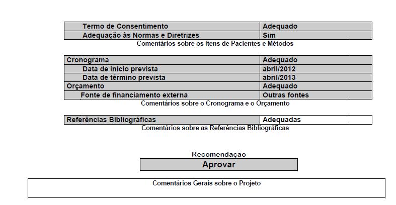
56 56

57 2.2 IRMANDADE SANTA CASA DE MISERICÓRDIA DE PORTO ALEGRE 57
Infecções virais no rim transplantado
 Infecções virais no rim transplantado Gabriel Teixeira Montezuma Sales Nefrologista Pós graduando em Nefrite INTRODUÇÃO Neoplasia DAE Perda do enxerto Infecção Viral Rejeição Mortalidade Cukuranovic et
Polioma vírus e Transplante Renal
 Polioma vírus e Transplante Renal Arthur Heinz Miehrig Médico Nefrologista UNIFESP/EPM! INTRODUÇÃO Polioma vírus é um pequeno vírus de DNA que infecta uma variedade de animais, incluindo humanos (1). Exemplos
UNIVERSIDADE PAULISTA CENTRO DE CONSULTORIA EDUCACIONAL DELANE CRISTINA DA SILVA AVALIAÇÃO CITOLÓGICA DO PAPILOMAVÍRUS HUMANO-HPV
 UNIVERSIDADE PAULISTA CENTRO DE CONSULTORIA EDUCACIONAL DELANE CRISTINA DA SILVA AVALIAÇÃO CITOLÓGICA DO PAPILOMAVÍRUS HUMANO-HPV RECIFE-PERNAMBUCO 2012 DELANE CRISTINA DA SILVA AVALIAÇÃO CITOLÓGICA DO
AVALIAÇÃO DA RESPOSTA IMUNE EM PACIENTES COM LEISHMANIOSE MUCOSA TRATADOS COM ANTIMONIAL
 RESUMO AVALIAÇÃO DA RESPOSTA IMUNE EM PACIENTES COM LEISHMANIOSE MUCOSA TRATADOS COM ANTIMONIAL PENTAVALENTE E PENTOXIFILINA Introdução: A leishmaniose mucosa (LM) é uma forma grave de apresentação da
Dissertação de mestrado
 Dissertação de mestrado Salvador (Bahia), 2017 Tratamento da leishmaniose cutânea com tamoxifeno e antimônio pentavalente: estudo piloto e análise in situ Camila Sampaio Ribeiro Professor-orientador: Paulo
Potencial da Telemedicina Dentária no diagnóstico oral infantil
 Potencial da Telemedicina Dentária no diagnóstico oral infantil ( RESUMO / ABSTRACT) Rui José de Oliveira Amável Morais 2010 Mestrado de Informática Médica Faculdade de Ciências Faculdade de Medicina Universidade
Introdução: O transplante renal é a melhor forma de substituição da função. renal em termos de esperança de vida e qualidade de vida.
 Resumo Introdução: O transplante renal é a melhor forma de substituição da função renal em termos de esperança de vida e qualidade de vida. As melhorias nas taxas de sobrevivência dos doentes e consequente
Prevalência de candidíase vulvovaginal e tricomoníase na citologia cérvicovaginal. corada por harris-shorr e sua associação com os sistemas de saúde
 AGENOR STORTI FILHO Prevalência de candidíase vulvovaginal e tricomoníase na citologia cérvico-vaginal corada por harris-shorr e sua associação com os sistemas de saúde pública e privada na região de Maringá-PR
UNIVERSIDADE FEDERAL DA BAHIA FACULDADE DE MEDICINA DA BAHIA PROGRAMA DE PÓS-GRADUAÇÃO EM CIÊNCIAS DA SAÚDE
 UNIVERSIDADE FEDERAL DA BAHIA FACULDADE DE MEDICINA DA BAHIA PROGRAMA DE PÓS-GRADUAÇÃO EM CIÊNCIAS DA SAÚDE CARVEL SUPRIEN CARACTERIZAÇÃO DAS MANIFESTAÇÕES CLÍNICAS, E RESPOSTA AO TRATAMENTO EM CRIANÇAS
Manifestações clínicas e resposta imune em uma coorte de indivíduos infectados pelo HTLV-1 com alta carga proviral e sem evidência de mielopatia
 1 Manifestações clínicas e resposta imune em uma coorte de indivíduos infectados pelo HTLV-1 com alta carga proviral e sem evidência de mielopatia I. RESUMO Introdução. A mielopatia associada ao vírus
Avaliação do Índice Apoptótico em Adenomas Pleomórficos de Glândulas Salivares
 PONTIFÍCIA UNIVERSIDADE CATÓLICA DE MINAS GERAIS Faculdade de Odontologia Avaliação do Índice Apoptótico em Adenomas Pleomórficos de Glândulas Salivares Paulo César de Lacerda Dantas Belo Horizonte- MG
Avaliação da telelaringoscopia no diagnóstico das lesões benignas da laringe
 Hospital do Servidor Público Municipal de São Paulo Avaliação da telelaringoscopia no diagnóstico das lesões benignas da laringe Márcio Cavalcante Salmito SÃO PAULO 2012 Márcio Cavalcante Salmito Avaliação
Influência das Variantes Genéticas Funcionais do Sistema Renina-Angiotensina na Doença Arterial Coronária.
 José Ramón Lanz Luces Influência das Variantes Genéticas Funcionais do Sistema Renina-Angiotensina na Doença Arterial Coronária. Tese apresentada à Faculdade de Medicina da Universidade de São Paulo para
Artigo Original TRATAMENTO DO CÂNCER DE CABEÇA E PESCOÇO NO IDOSO ACIMA DE 80 ANOS
 Artigo Original TRATAMENTO DO CÂNCER DE CABEÇA E PESCOÇO NO IDOSO ACIMA DE 80 ANOS HEAD AND NECK CANCER TREATMENT IN ELDERLY PATIENTS OVER 80 YEARS OLD 1,4,6 TERENCE PIRES DE FARIAS 5 GABRIEL MANFRO 1,2,3
INVESTIGAÇÃO FARMACOEPIDEMIOLÓGICA DO USO DO CLONAZEPAM NO DISTRITO SANITÁRIO LESTE EM NATAL-RN
 RODRIGO DOS SANTOS DINIZ DISSERTAÇÃO A SER APRESENTADA AO PROGRAMA DE PÓS-GRADUAÇÃO EM CIÊNCIAS DA SAÚDE, COMO PARTE DOS REQUISITOS PARA A OBTENÇÃO DO TÍTULO DE MESTRE. Rodrigo dos Santos Diniz Orientadora:
Questionário - Proficiência Clínica
 Questionário - Proficiência Clínica DISMORFISMO ERITROCITÁRIO Elaborador José Antonio Tesser Poloni. Mestre em Ciências da Saúde (UFCSPA), Farmacêutico-Bioquímico (PUCRS), Bioquímico responsável pelo setor
RESUMO ANÁLISE DO TROPISMO DE LEISHMANIA BRAZILIENSIS
 RESUMO ANÁLISE DO TROPISMO DE LEISHMANIA BRAZILIENSIS PARA A MUCOSA NASAL ANTES E LONGO PERÍODO APÓS O TRATAMENTO DALEISHMANIOSE LOCALIZADA. INTRODUÇÃO: Leishmania sp. pode estar presente na mucosa nasal
Glomerulonefrite Membranosa Idiopática: um estudo de caso
 Glomerulonefrite Membranosa Idiopática: um estudo de caso Cláudia Maria Teixeira de Carvalho Dissertação de Mestrado Integrado em Medicina Faculdade de Ciências da Saúde Universidade da Beira Interior
Casos Clínicos Sessão 5. Caso 6
 Casos Clínicos Sessão 5. Caso 6 Receptor: S.A.V, 45 anos, masculino, branco. DRC por DM1 HD por 1 ano e 2 meses. Tx renal há 1 ano e 6 meses Outras comorbidades: HAS. Doador: Doador falecido - F.G.H, 50
Avaliação do padrão e número de sítios de fosforilação (EPIYA) da proteína CagA de H. pylori e risco de carcinoma gástrico e úlcera duodenal
 da proteína CagA de H. pylori e risco de carcinoma gástrico e úlcera duodenal") UNIVERSIDADE FEDERAL DE MINAS GERAIS INSTITUTO DE CIÊNCIAS BIOLÓGICAS CURSO DE PÓS-GRADUAÇÃO EM MICROBIOLOGIA Avaliação do padrão e número de sítios de fosforilação (EPIYA) da proteína CagA de H. pylori
UNIVERSIDADE FEDERAL DE MINAS GERAIS INSTITUTO DE CIÊNCIAS BIOLÓGICAS CURSO DE PÓS-GRADUAÇÃO EM MICROBIOLOGIA Avaliação do padrão e número de sítios de fosforilação (EPIYA) da proteína CagA de H. pylori
INTRODUÇÃO LESÃO RENAL AGUDA
 INTRODUÇÃO Pacientes em tratamento imunossupressor com inibidores de calcineurina estão sob risco elevado de desenvolvimento de lesão, tanto aguda quanto crônica. A manifestação da injuria renal pode se
ESTUDO DA FREQÜÊNCIA DE PARASITAS E DE OVOS NAS CAVIDADES APENDICULARES DAS APENDICITES AGUDAS *
 ESTUDO DA FREQÜÊNCIA DE PARASITAS E DE OVOS NAS CAVIDADES APENDICULARES DAS APENDICITES AGUDAS * DAOIZ MENDOZA** HENRI CHAPLIN RIVOIRE*** MARCELO DORNELES DE SOUSA**** RESUMO Os autores fizeram um estudo
Qual o papel do Teste HPV- DNA no rastreamento do câncer do colo uterino? Victor Hugo Melo
 Qual o papel do Teste HPV- DNA no rastreamento do câncer do colo uterino? Victor Hugo Melo Uso da biologia molecular no rastreamento do câncer de colo uterino Victor Hugo Melo Faculdade de Medicina da
INVESTIGAÇÃO CLÍNICO-LABORATORIAL DE INFECÇÃO POR POLIOMAVÍRUS EM TRANSPLANTE RENAL
 488 INVESTIGAÇÃO CLÍNICO-LABORATORIAL DE INFECÇÃO POR POLIOMAVÍRUS EM TRANSPLANTE RENAL Polyomavirus infection in kidney transplantation Juliana Montagner, Tatiana Michelon, Regina Schroeder, Alexandre
Atividade, Gravidade e Prognóstico de pacientes com Lúpus Eritematoso Sistêmico antes, durante e após prima internação
 doi: 10.12662/2317-3076jhbs.v2i2.56.p.65.2014 ARTIGO DE REVISÃO Atividade, Gravidade e Prognóstico de pacientes com Lúpus Eritematoso Sistêmico antes, durante e após prima internação Systemic Lupus Erythematosus
Implementation of BE requirements: Brazilian Experience
 13 th ICDRA Implementation of BE requirements: Brazilian Experience RODRIGO CRISTOFOLETTI Head of Department of Bioequivalence Brazilian National Health Surveillance Agency (ANVISA) Historical view Future
Pró-Reitoria Acadêmica Escola de Saúde e Medicina Programa de Pós-Graduação Stricto Sensu em Gerontologia
 Pró-Reitoria Acadêmica Escola de Saúde e Medicina Programa de Pós-Graduação Stricto Sensu em Gerontologia ESTUDO DA COBERTURA VACINAL CONTRA INFLUENZA NO PERFIL DE MORTALIDADE DE IDOSOS NO BRASIL Autora:
JULIANA T. DOMINGUES 1 FERNANDA DE PARIS 2 VIRGÍNIA M. SCHMITT 3
 ARTIGO ORIGINAL Proposta de reação em cadeia da polimerase (PCR) semi-nested para pesquisa e diferenciação dos vírus BK e JC Proposal of semi-nested polimerase chain reaction (PCR) for diagnosing and differentiating
Lucas de Assis Soares, Luisa Nunes Ramaldes, Taciana Toledo de Almeida Albuquerque, Neyval Costa Reis Junior. São Paulo, 2013
 COMPARATIVE STUDY OF THE ATMOSPHERIC DISPERSION MODELS AND THROUGH THE ANALYSIS OF AIR QUALITY IN THE METROPOLITAN REGION OF GRANDE VITÓRIA Lucas de Assis Soares, Luisa Nunes Ramaldes, Taciana Toledo de
IDENTIFICAÇÃO E QUANTIFICAÇÃO DE MICRORGANISMOS EM INFECÇÕES PERIAPICAIS CRÔNICAS PÓS-TERAPIA ENDODÔNTICA
 UNIVERSIDADE FEDERAL DO ESPÍRITO SANTO CENTRO DE CIÊNCIAS DA SAÚDE PROGRAMA DE PÓS GRADUAÇÃO EM CLÍNICA ODONTOLÓGICA MESTRADO PROFISSIONAL EM CLÍNICA ODONTOLÓGICA WEDERSON TAVARES FURTADO IDENTIFICAÇÃO
Caso Clínico. (abordagem de Glomerulopatias pós-transplante)
") 16 A 18 DE ABRIL, 2018 HOSPITAL DO RIM, SÃO PAULO, SP Caso Clínico (abordagem de Glomerulopatias pós-transplante) Dra. Maria Almerinda V. F. Ribeiro Alves Dr. Henrique Machado Proença Caso clínico Transplante
16 A 18 DE ABRIL, 2018 HOSPITAL DO RIM, SÃO PAULO, SP Caso Clínico (abordagem de Glomerulopatias pós-transplante) Dra. Maria Almerinda V. F. Ribeiro Alves Dr. Henrique Machado Proença Caso clínico Transplante
GESF no Transplante. Introdução
 GESF no Transplante Introdução A glomeruloesclerose segmentar e focal (GESF) é um padrão histológico de lesão renal que pode apresentar diferentes etiologias, incluindo doenças imunológicas, genéticas,
PREVENÇÃO DE CÂNCER DE COLO UTERINO: CONCEPÇÃO DOS ENFERMEIROS DAS UNIDADES BÁSICAS DE SAÚDE DE MOSSORÓ/RN
 FACULDADE DE ENFERMAGEM NOVA ESPERANÇA DE MOSSORÓ FACENE/RN ÉRICA DANIELY OLIVEIRA DA COSTA PREVENÇÃO DE CÂNCER DE COLO UTERINO: CONCEPÇÃO DOS ENFERMEIROS DAS UNIDADES BÁSICAS DE SAÚDE DE MOSSORÓ/RN MOSSORÓ
Pró-Reitoria Acadêmica Escola de Exatas, Arquitetura e Meio Ambiente. Curso de Ciências Biológicas. Curso de Ciências Biológicas
 Pró-Reitoria Acadêmica Escola de Exatas, Arquitetura e Meio Ambiente Curso de Ciências Biológicas Trabalho Pró-Reitoria de Conclusão de Graduação de Curso Curso de Ciências Biológicas Projeto de Trabalho
CARACTERIZAÇÃO CLÍNICA E RADIOLÓGICA DA ARTROPATIA ASOCIADA AO HTLV-1
 CARACTERIZAÇÃO CLÍNICA E RADIOLÓGICA DA ARTROPATIA ASOCIADA AO HTLV-1 RESUMO Introdução: No passado doenças reumáticas sistêmicas foram identificadas como sendo mais frequentes em indivíduos infectados
Coeficiente kappa: Mede a concordância entre dois observadores, métodos ou instrumentos, considerando variáveis qualitativas nominais.
 Concordância Coeficiente kappa: Mede a concordância entre dois observadores, métodos ou instrumentos, considerando variáveis qualitativas nominais. Coeficiente kappa ponderado: Mede a concordância entre
Detecção quantitativa de vírus BK em receptores de transplante renal: um estudo prospectivo de validação
 Artigo Original Original Article Detecção quantitativa de vírus BK em receptores de transplante renal: um estudo prospectivo de validação Quantitative detection of BK virus in kidney transplant recipients:
Avaliação crítica de artigos sobre diagnóstico
 Avaliação crítica de artigos sobre diagnóstico Objetivos da aula Formulação da pergunta Identificação e seleção do estudo Roteiro de avaliação da qualidade de um artigo sobre diagnóstico Sintetizando informações
Teste do Tempo & Troco
 Instituto Superior Miguel Torga Escola Superior de Altos Estudos Teste do Tempo & Troco Estudo de Validação Edgar Correia Dissertação de Mestrado em Psicologia Clínica Coimbra, 2011 Teste do Tempo & Troco
ELVIANI BASSO MOURA AVALIAÇÃO DA PREDISPOSIÇÃO E IMPACTO CLÍNICO DA REPLICAÇÃO DO VÍRUS BK EM TRANSPLANTADOS RENAIS
 ELVIANI BASSO MOURA AVALIAÇÃO DA PREDISPOSIÇÃO E IMPACTO CLÍNICO DA REPLICAÇÃO DO VÍRUS BK EM TRANSPLANTADOS RENAIS JOINVILLE 2016 1 ELVIANI BASSO MOURA AVALIAÇÃO DA PREDISPOSIÇÃO E IMPACTO CLÍNICO DA
UNIVERSIDADE REGIONAL INTEGRADA DO ALTO URUGUAI E DAS MISSÕES CAMPUS DE ERECHIM DEPARTAMENTO DE CIÊNCIAS DA SAÚDE CURSO DE ENFERMAGEM
 UNIVERSIDADE REGIONAL INTEGRADA DO ALTO URUGUAI E DAS MISSÕES CAMPUS DE ERECHIM DEPARTAMENTO DE CIÊNCIAS DA SAÚDE CURSO DE ENFERMAGEM ESTRATIFICAÇÃO DE RISCO CARDIOVASCULAR E PERDA DA SENSIBILIDADE DOS
ANABELA VIRGÍNIA DOS SANTOS FLORES DA ROCHA ESTIMAÇÃO ROBUSTA EM MODELOS LINEARES DE EQUAÇÕES SIMULTÂNEAS
 Universidade de Aveiro Departamento de Matemática 2010 ANABELA VIRGÍNIA DOS SANTOS FLORES DA ROCHA ESTIMAÇÃO ROBUSTA EM MODELOS LINEARES DE EQUAÇÕES SIMULTÂNEAS Universidade de Aveiro Departamento de Matemática
Escola Superior de Altos Estudos
 Escola Superior de Altos Estudos Defeito cognitivo, sintomas de depressão e satisfação com a vida em idosos sob resposta social do concelho de Coimbra INÊS TORRES PENA Dissertação Apresentada ao ISMT para
Instrução Normativa nº 10, de 19/12/2018
 Instrução Normativa nº 10, de 19/12/2018 Dispõe sobre a participação não presencial de membros de Comissões Examinadoras (Bancas) do Programa de Pós-Graduação em Ecologia e Biodiversidade. Artigo 1º Conforme
COMPARAÇÃO DE DOIS MÉTODOS DE DIAGNÓSTICO DE Salmonella Enteritidis. COMPARISON OF TWO DIAGNOSTIC METHODS OF Salmonella Enteritidis
 1 COMPARAÇÃO DE DOIS MÉTODOS DE DIAGNÓSTICO DE Salmonella Enteritidis COMPARISON OF TWO DIAGNOSTIC METHODS OF Salmonella Enteritidis SIMONE ROSA DIDONÉ 1, CAROLINA DE OLIVEIRA FREITAS 1, AMANDA FERNANDES
ANÁLISE DA GENOTOXICIDADE DAS ÁGUAS DA LAGOA DE EXTREMOZ-RN
 UNIVERSIDADE FEDERAL DO RIO GRANDE DO NORTE CENTRO DE BIOCIÊNCIAS DEPARTAMENTO DE BIOLOGIA CELULAR E GENÉTICA PROGRAMA DE PÓS-GRADUAÇÃO EM GENÉTICA E BIOLOGIA MOLECULAR ANÁLISE DA GENOTOXICIDADE DAS ÁGUAS
Retrovírus Felinos. Fernando Finoketti
 Retrovírus Felinos Fernando Finoketti Porto Alegre, Rio Grande do Sul, Brasil Maio de 2014 Retrovírus - Características Capsídeo icosaédrico. Possuem envelope. Genoma composto de duas moléculas idênticas
LINA MONETTA ANÁLISE EVOLUTIVA DO PROCESSO DE CICATRIZAÇÃO EM ÚLCERAS DIABÉTICAS, DE PRESSÃO E VENOSAS COM USO DE PAPAÍNA
 LINA MONETTA ANÁLISE EVOLUTIVA DO PROCESSO DE CICATRIZAÇÃO EM ÚLCERAS DIABÉTICAS, DE PRESSÃO E VENOSAS COM USO DE PAPAÍNA Dissertação de Mestrado apresentada a Escola de Enfermagem da Universidade de São
Detecção precoce de sepse e segurança do paciente. O que fazer? Flavia Machado
 Detecção precoce de sepse e segurança do paciente. O que fazer? Flavia Machado O que fazer? World Health Assembly urges member states to: Develop national policy and processes to improve the prevention,
A Tool to Evaluate Stuck-Open Faults in CMOS Logic Gates
 FURG A Tool to Evaluate Stuck-Open Faults in CMOS Logic Gates Alexandra L. Zimpeck, Cristina Meinhardt e Paulo F. Butzen Summary Introduction Motivation Stuck-Open Faults Stuck-Open Faults in Nanometer
OFERTA DE DISCIPLINAS PARA 2014/2. Obs: Não serão aceitos campos a definir
 Disciplina Código Ementa (campo obrigatório apenas para Tópicos) Topicos especiais Em Biologia Celular I - Analises em Citometria de fluxo* Em Biologia Celular II Biologia de linfócitos T em condições
Prof. Dr. Marcelo Nascimento Burattini
 Mathematical models of Hepatitis C and its Consequences in Public Health mnburatt@usp.br Hepatitis C virus first described in 1988 mnburatt@usp.br Estudos de soroprevalencia e modelos dinâmicos em dengue
I. RESUMO TROMBOEMBOLISMO VENOSO APÓS O TRANSPLANTE PULMONAR EM ADULTOS: UM EVENTO FREQUENTE E ASSOCIADO A UMA SOBREVIDA REDUZIDA.
 I. RESUMO TROMBOEMBOLISMO VENOSO APÓS O TRANSPLANTE PULMONAR EM ADULTOS: UM EVENTO FREQUENTE E ASSOCIADO A UMA SOBREVIDA REDUZIDA. Introdução: A incidência de tromboembolismo venoso (TEV) após o transplante
PAULA CRISTINA MONTEIRO DA FONSECA VALE POLIOMAVIRUS BK: COMPARAÇÃO ENTRE CITOLOGIA E HIBRIDIZAÇÃO IN SITU
 Universidade de Aveiro Departamento de Biologia 2009 PAULA CRISTINA MONTEIRO DA FONSECA VALE POLIOMAVIRUS BK: COMPARAÇÃO ENTRE CITOLOGIA E HIBRIDIZAÇÃO IN SITU Universidade de Aveiro Departamento de Biologia
Service quality in restaurants: an experimental analysis performed in Brazil
 . XIII INTERNATIONAL CONFERENCE ON INDUSTRIAL ENGINEERING AND OPERATIONS MANAGEMENT Energy that moves production: a dialogue among integration, project and sustainability 09-11 October 2007 Service quality
Universidade de São Paulo
 Universidade de São Paulo Faculdade de Medicina de Ribeirão Preto Departamento de Medicina Social Maria Angélica de Figueiredo Campos PERCEPÇÃO E AVALIAÇÃO DOS ALUNOS DO CURSO DE MEDICINA DE UMA ESCOLA
Artigo Original TUMORES DO PALATO DURO: ANÁLISE DE 130 CASOS HARD PALATE TUMORS: ANALISYS OF 130 CASES ANTONIO AZOUBEL ANTUNES 2
 Artigo Original TUMORES DO PALATO DURO: ANÁLISE DE 130 CASOS HARD PALATE TUMORS: ANALISYS OF 130 CASES 1 ANTONIO AZOUBEL ANTUNES 2 ANTONIO PESSOA ANTUNES 3 POLLIANA VILAÇA SILVA RESUMO Introdução: O câncer
NEFROTOXICIDADE MEDICAMENTOSA. Dr. Carlos Alberto Balda Professor afiliado da Disciplina de Nefrologia da EPM - UNIFESP
 NEFROTOXICIDADE MEDICAMENTOSA Dr. Carlos Alberto Balda Professor afiliado da Disciplina de Nefrologia da EPM - UNIFESP FUNÇÃO RENAL 7.507 PACIENTES INTERCONSULTA HSP/ UNIFESP NORMAL; 392 AGUDO; 3785
5.1. Prevalência de anticorpos anti-hhv-8 em doadores de
 RESULTADOS 67 5. RESULTADOS 5.1. Prevalência de anticorpos anti-hhv-8 em doadores de sangue Analisamos 400 doadores de sangue entre outubro de 2000 a agosto de 2001. Foram detectados anticorpos em 16 (4%)
Seguimento após tratamento das Neoplasias Intraepiteliais Cervicais. Fábio Russomano --IFF/Fiocruz Maio de de 2014
 Seguimento após tratamento das Neoplasias Intraepiteliais Cervicais Fábio Russomano --IFF/Fiocruz Maio de de 2014 Possíveis conflitos de interesses Responsável por serviço público de Patologia Cervical
Após um episódio de ITU, há uma chance de aproximadamente 19% de aparecimento de cicatriz renal
 Compartilhe conhecimento: Devemos ou não continuar prescrevendo antibiótico profilático após diagnóstico da primeira infecção urinária? Analisamos recente revisão sistemática e trazemos a resposta. Em
AGENESIA DO SEGUNDO PRÉ-MOLAR INFERIOR: AVALIAÇÃO OCLUSAL E RADICULAR DO CORRESPONDENTE DECÍDUO
 INSTITUTO SUPERIOR DE CIÊNCIAS DA SAÚDE NORTE DISSERTAÇÃO PARA OBTENÇÃO DO GRAU DE MESTRE EM ORTODONTIA AGENESIA DO SEGUNDO PRÉ-MOLAR INFERIOR: AVALIAÇÃO OCLUSAL E RADICULAR DO CORRESPONDENTE DECÍDUO Ana
Delineamento de estudo caso-controle
 Delineamento de estudo caso-controle EXPOSTOS EXPOSIÇÃO AO FATOR DE RISCO CASOS/CONTROLES POPULAÇÃO SIM NÃO SIM NÃO TEMPO PESQUISA CASOS (Apresentam a doença) CONTROLES (Não apresentam a doença) Estimativa
Métodos de Pesquisa e Diagnóstico dos Vírus
 Métodos de Pesquisa e Diagnóstico dos Vírus Estratégias Isolamento em sistemas vivos Pesquisa de antígeno viral Pesquisa de anticorpos Pesquisa do ácido nucléico viral (DNA ou RNA) Pré requisitos para
Instituto Politécnico de Tomar. Controlo de TCA e outros off-flavours na Cortiça. Relatório de Estágio. Ana Leonor da Costa Vila Mendes
 Instituto Politécnico de Tomar Escola Superior de Tecnologia de Tomar Controlo de TCA e outros off-flavours na Cortiça Relatório de Estágio Ana Leonor da Costa Vila Mendes Mestrado em Tecnologia Química
Imunologia da infecção pelo HIV
 Imunologia da infecção pelo HIV Histórico e epidemiologia 1981 - Em oito meses foram diagnosticados 5 casos de pneumonia pelo FUNGO (Pneumocystis jiroveci). Entre 1967 e 1979 foram diagnosticados apenas
Assessment of minimum detection limit of Nested-PCR
 Artigo Original Determinação do limite mínimo de detecção da técnica de Nested-PCR para os poliomavírus JC e BK Assessment of minimum detection limit of Nested-PCR for JC and BK polyomaviruses Aline dos
Camila Ap. Marques Faria de Melo Kíssila Brito Fiszer
 Camila Ap. Marques Faria de Melo Kíssila Brito Fiszer EFEITO DA FACILITAÇÃO NEUROMUSCULAR PROPRIOCEPTIVA (KABAT) SOBRE PARÂMETROS RESPIRATÓRIOS E EMOCIONAIS EM PACIENTE PORTADOR DE DOENÇA PULMONAR OBSTRUTIVA
Portuguese registry on congenital CMV infection Preliminary results
 Portuguese registry on congenital CMV infection Preliminary results Paulo Paixão, Maria Teresa Neto, Maria João Brito, Graça Rocha, Teresa Marques Portuguese Paediatric Surveillance Unit Faculdade de Ciências
Programa de Acesso Precoce e de Partilha de Risco em Oncologia. Ricardo da Luz Centro Hospitalar Lisboa Central
 Programa de Acesso Precoce e de Partilha de Risco em Oncologia Ricardo da Luz Centro Hospitalar Lisboa Central Programas de Acesso Precoce Diário de Notícias 3 de Dezembro 20 500 doentes em estado grave
artus BK Virus QS-RGQ Kit
 artus BK Virus QS-RGQ Kit Características de desempenho artus BK Virus QS-RGQ Kit, versão 1, 4514363 Verificar a disponibilidade de novas revisões de rotulagem eletrónica em www.qiagen.com/products/artusbkvirusrgpcrkit.aspx
EFEITO DA ESTIMULAÇÃO VESTIBULAR NO PERFIL SENSORIAL DE CRIANÇAS COM PARALISIA CEREBRAL: ENSAIO CLÍNICO RANDOMIZADO ALEATORIZADO
 1 UNIVERSIDADE DE BRASÍLIA-UnB FACULDADE DE CEILÂNDIA-FCE CURSO DE FISIOTERAPIA YASMIN SANTANA MAGALHÃES EFEITO DA ESTIMULAÇÃO VESTIBULAR NO PERFIL SENSORIAL DE CRIANÇAS COM PARALISIA CEREBRAL: ENSAIO
LEONORA ZOZULA BLIND POPE. AVALIAÇÃO DO PERFIL EPIDEMIOLÓGICO E DA PROTEINA p 53 EM 12 GLIOBLASTOMAS PEDIÁTRICOS.
 LEONORA ZOZULA BLIND POPE AVALIAÇÃO DO PERFIL EPIDEMIOLÓGICO E DA PROTEINA p 53 EM 12 GLIOBLASTOMAS PEDIÁTRICOS. Dissertação apresentada como requisito parcial à obtenção do grau de Mestre. Programa de
COLONIZAÇÃO BACTERIANA NA CAVIDADE BUCAL POR PATÓGENOS CAUSADORES DA PNEUMONIA ASSOCIADA À
 UNIVERSIDADE FEDERAL DO TOCANTINS PRÓ-REITORIA DEPESQUISA E PÓS-GRADUAÇÃO PROGRAMA DE PÓS-GRADUAÇÃO EM CIÊNCIAS DA SAÚDE FREDERICO EUGÊNIO COLONIZAÇÃO BACTERIANA NA CAVIDADE BUCAL POR PATÓGENOS CAUSADORES
PONTIFÍCIA UNIVERSIDADE CATÓLICA DO RIO GRANDE DO SUL FACULDADE DE ODONTOLOGIA PROGRAMA DE PÓS-GRADUAÇÃO EM ODONTOLOGIA DOUTORADO EM PRÓTESE DENTÁRIA
 PONTIFÍCIA UNIVERSIDADE CATÓLICA DO RIO GRANDE DO SUL FACULDADE DE ODONTOLOGIA PROGRAMA DE PÓS-GRADUAÇÃO EM ODONTOLOGIA DOUTORADO EM PRÓTESE DENTÁRIA DANIELA DISCONZI SEITENFUS REHM PREVALÊNCIA DE DIFERENTES
Comparação da contagem de plaquetas entre o método de Fônio e automação
 Comparação da contagem de plaquetas entre o método de Fônio e automação Comparison of the platelet count between the Fonio method and automation Avila FA a, Oliveira LS a a: Faculdades Metropolitanas Unidas
APLICAÇÕES DA BIOLOGIA MOLECULAR NO DIAGNÓSTICO E TRATAMENTO DA HEPATITE B. Maio
 APLICAÇÕES DA BIOLOGIA MOLECULAR NO DIAGNÓSTICO E TRATAMENTO DA HEPATITE B Maio - 2009 BIOLOGIA MOLECULAR & HEPATITE B INFECÇÃO CRÔNICA PELO VÍRUS B Afeta mais de 300 milhões de indivíduos em todo o mundo.
Índice. Índice... ii Sumário... iii Abstract... v
 2ª ed Caracterização de reacções adversas a medicamentos notificadas à Unidade de Farmacovigilância do Norte pelo Serviço de Imunoalergologia do Centro Hospitalar de São João do Porto Maria João Baldaia
RECURSOS EXISTENTES DE PESSOAL MÉDICO E PARA-MÉDICO NA REGIÃO DO GRANDE SÃO PAULO EM 1966
 RECURSOS EXISTENTES DE PESSOAL MÉDICO E PARA-MÉDICO NA REGIÃO DO GRANDE SÃO PAULO EM 1966 João YUNES (1) YUNES, J. Recursos existentes de pessoal médico e para-médico na região do Grande São Paulo (Brasil)
Doença Residual Mínima (DRM) e Transplante de Células Tronco Hematopoéticas (TCTH)
 e Transplante de Células Tronco Hematopoéticas (TCTH)") Doença Residual Mínima (DRM) e Transplante de Células Tronco Hematopoéticas (TCTH) How and why minimal residual disease studies are necessary in leukemia (LeukemiaNet, Marie C. Bene, haematologica, 2009):
Doença Residual Mínima (DRM) e Transplante de Células Tronco Hematopoéticas (TCTH) How and why minimal residual disease studies are necessary in leukemia (LeukemiaNet, Marie C. Bene, haematologica, 2009):
Ferramentas de Apoio à Decisão Clínica em MGF
 Ferramentas de Apoio à Decisão Clínica em MGF Ana Resende Mateus MF/OF USF FF-mais, ACES Almada-Seixal CIMGFLVT 2015 Curso A Consulta CIMGF-LVT Ferramentas de apoio à decisão clínica O que são? Para que
Revisão Sistemática: Conceitos Básicos
 SSC5905 Revisão Sistemática em Engenharia de Software Revisão Sistemática: Conceitos Básicos Profa. Dra. Elisa Yumi Nakagawa elisa@icmc.usp.br ICMC/USP São Carlos 1. Semestre 2013 Questões preliminares
Lesão intraepitelial de baixo grau: como seguir? Fábio Russomano IFF/Fiocruz Paranacolpo 25 a 27 de julho de 2013
 Lesão intraepitelial de baixo grau: como seguir? Fábio Russomano IFF/Fiocruz Paranacolpo 25 a 27 de julho de 2013 Qual o objetivo de seguir uma lesão de baixo grau? a. Verificar se regrediu? b. Verificar
LISTA DE TABELAS. Página
 vi LISTA DE TABELAS Página TABELA 1 - Tipos psicológicos da equipe A 66 TABELA 2 - Tipos psicológicos da equipe B 66 TABELA 3 - Média dos estados de humor da equipe A em quatro 70 momentos diferentes TABELA
03/07/2012. DIAGNÓSTICO Avanços DIAGNÓSTICO. Avanços no Diagnóstico da Tuberculose. Encontro Nacional de Infecções Respiratórias e Tuberculose
 Avanços no Diagnóstico da Tuberculose Encontro Nacional de Infecções Respiratórias e Tuberculose Fernanda C Q Mello Instituto de Doenças do Tórax Faculdade de Medicina - UFRJ fcqmello@hucff.ufrj.br Reduzir
Aos meus alunos e amigos.
 ii Aos meus alunos e amigos. iii Agradeço especialmente ao professor Edgard Bruno Cornachione Júnior por ter se mostrado sempre presente em todos os momentos desta pesquisa, como orientador e amigo. Por
Agenda. Cost-effectiveness analysis of vemurafenib compared to dacarbazine for the metastatic melanoma treatment in Brazilian Public Health System
 Cost-effectiveness analysis of vemurafenib compared to dacarbazine for the metastatic melanoma treatment in Brazilian Public Health System Telma Rodrigues Caldeira Priscila Louly Gabrielle Troncoso Misani
Declaração de Conflitos de Interesse
 Declaração de Conflitos de Interesse Nada a ser declarado. Neoplasias de Sistema Nervoso Central Diagnóstico laboratorial do comprometimento do Sistema Nervoso Central por Linfomas Não Hodgkin Alvaro Rodrigues
INDICADORES BIOQUÍMICOS DA FUNÇÃO RENAL EM IDOSOS
 1 ÁREA TEMÁTICA: ( ) COMUNICAÇÃO ( ) CULTURA ( ) DIREITOS HUMANOS E JUSTIÇA ( ) EDUCAÇÃO ( ) MEIO AMBIENTE ( x ) SAÚDE ( ) TECNOLOGIA E PRODUÇÃO ( ) TRABALHO INDICADORES BIOQUÍMICOS DA FUNÇÃO RENAL EM
TRATAMENTO PREEMPTIVO NA HEPATITE B: QUEM DEVERIA SER TRATADO?
 TRATAMENTO PREEMPTIVO NA HEPATITE B: QUEM DEVERIA SER TRATADO? WORNEI BRAGA FMT-HVD MANAUS - BRASIL VHB E SUA IMPORTÂNCIA VHB E SUA IMPORTÂNCIA VÍRUS DNA PEQUENO CIRCULAR FITA DUPLA PARCIAL Hepadnaviridae
Infecção por Poliomavírus: tratamento com Cidofovir e Imunoglobulina Inespecífica, um esquema promissor?
 INFECÇÃO POR POLIOMAVÍRUS: TRATAMENTO COM CIDOFOVIR E IMUNOGLOBULINA INESPECÍFICA, UM ESQUEMA PROMISSOR? Case Report Rev Port Nefrol Hipert 2006; 20 (1): 51-60 Infecção por Poliomavírus: tratamento com
SUMÁRIO SUMÁRIO... LISTA DE FIGURAS... LISTA DE TABELAS... ÍNDICE DE ANEXOS... RESUMO... ABSTRACT INTRODUÇÃO... 1
 SUMÁRIO Página SUMÁRIO... LISTA DE FIGURAS... LISTA DE TABELAS... ÍNDICE DE ANEXOS... i iii iv iv RESUMO... ABSTRACT... v vii 1. INTRODUÇÃO... 1 1.1 Métodos para detecção de E. sakazakii... 20 2. OBJETIVOS...
Poliomavírus um patógeno emergente para receptores de transplantes
 184 Montagner JM, Michelon TF, Schroeder RB, Fontanelle BT, Oliveira ATD, Silveira JG, Graudenz MS, Alexandre COP, Neumann J avanços médicos Poliomavírus um patógeno emergente para receptores de transplantes
VACCINATION PLAN FOR AVIAN INFLUENZA IN A GAME BIRD HOLDING OF BREEDING MALLARD DUCKS (Anas platyrhynchus) PORTUGAL
 PORTUGAL") PAFF Com Brussels, 18-19 September 2017 VACCINATION PLAN FOR AVIAN INFLUENZA IN A GAME BIRD HOLDING OF BREEDING MALLARD DUCKS (Anas platyrhynchus) PORTUGAL www.dgav.pt History Outbreak of LPAI subtype
PAFF Com Brussels, 18-19 September 2017 VACCINATION PLAN FOR AVIAN INFLUENZA IN A GAME BIRD HOLDING OF BREEDING MALLARD DUCKS (Anas platyrhynchus) PORTUGAL www.dgav.pt History Outbreak of LPAI subtype
CENTRO DE INFORMAÇÃO SOBRE MEDICAMENTOS - CIM: AVALIAÇÃO DA INFORMAÇÃO PRESTADA VIVIANE DO NASCIMENTO E SILVA
 CENTRO DE INFORMAÇÃO SOBRE MEDICAMENTOS - CIM: AVALIAÇÃO DA INFORMAÇÃO PRESTADA VIVIANE DO NASCIMENTO E SILVA CENTRO DE INFORMAÇÃO SOBRE MEDICAMENTOS CIM: AVALIAÇÃO DA INFORMAÇÃO PRESTADA DISSERTAÇÃO APRESENTADA
VGM. VGM information. ALIANÇA VGM WEB PORTAL USER GUIDE June 2016
 Overview The Aliança VGM Web portal is an application that enables you to submit VGM information directly to Aliança via our e-portal Web page. You can choose to enter VGM information directly, or to download
ROBERTA RIBEIRO SILVA. CRESCIMENTO DE Salmonella enteritidis var. Typhimurium EM DIETAS ENTERAIS
 ROBERTA RIBEIRO SILVA CRESCIMENTO DE Salmonella enteritidis var. Typhimurium EM DIETAS ENTERAIS Tese apresentada à Universidade Federal de Viçosa, como parte das exigências do Programa de Pós- Graduação
AS FUNÇÕES DA INDENIZAÇÃO POR DANOS MORAIS E A PREVENÇÃO DE DANOS FUTUROS
 MARCELA ALCAZAS BASSAN AS FUNÇÕES DA INDENIZAÇÃO POR DANOS MORAIS E A PREVENÇÃO DE DANOS FUTUROS Dissertação de Mestrado Apresentada ao Departamento de Direito Civil Orientador Prof. Titular Rui Geraldo
AVALIAÇÃO DO DESEMPENHO DA TÉCNICA DE PCR EM TEMPO REAL EM DIFERENTES AMOSTRAS BIOLÓGICAS UTILIZADAS PARA O DIAGNÓSTICO DA TUBERCULOSE EXTRAPULMONAR
 AVALIAÇÃO DO DESEMPENHO DA TÉCNICA DE PCR EM TEMPO REAL EM DIFERENTES AMOSTRAS BIOLÓGICAS UTILIZADAS PARA O DIAGNÓSTICO DA TUBERCULOSE EXTRAPULMONAR Autor: Fabiana Cristina Fulco Santos 1 ; Coautor: Luanna
AVALIAÇÃO DA FUNÇÃO RENAL EM DIABÉTICOS ADULTOS*
 AVALIAÇÃO DA FUNÇÃO RENAL EM DIABÉTICOS ADULTOS* BRAGA, Ana Karolina Paiva 1 ; PEREIRA, Edna Regina Silva 2, NAGHETTINI, Alessandra Vitorino 3, BATISTA, Sandro Rogério Rodrigues 4 Palavras-chave: doença